experiment · Nature communications · la publicación, 20 may 2026 · gratis
Una inteligencia artificial ayudó a leer ecografías de mama en China y mejoró la precisión de los radiólogos
El estudio se hizo en hospitales chinos. Fuera de ese país, sus autores dicen que la herramienta todavía necesita más pruebas.
Versión breve · la versión detallada sigue, unos 6 min
Pregunte al chat de weeklyAI
Pregúnteme por este estudio: a quiénes se estudió, qué encontró y qué no dice.
Las conversaciones se guardan mientras exista weeklyAI, para mejorar la publicación. Se responde en el idioma en que usted escribe.
- El estudio, de un vistazo
- Quiénes
- Mujeres a las que se les hizo una ecografía de mama
- Cuántos
- 6,817
- Dónde
- China
- Cuándo
- No lo dice el pasaje
- Tipo de estudio
- experiment
- Quién lo hizo
- Investigadores de hospitales y universidades de China
- El límite que importa
- Solo se siguió de cerca al grupo de riesgo alto, y los autores piden más pruebas fuera de China.
Radiólogos leyendo solos frente a radiólogos con ayuda del programa
Su precisión pasó de 0.687 a 0.736
Su precisión mejoró
Si a usted le hacen una ecografía de mama, puede preguntar si el centro usa un programa de inteligencia artificial como segunda lectura.
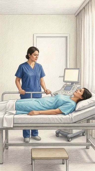
Investigadores en China construyeron un programa de computadora llamado BIRD. El programa lee imágenes de ecografía de mama y clasifica cada lesión según la escala BI-RADS, que ordena los hallazgos por nivel de sospecha.
En su propia prueba interna, BIRD clasificó bien 84 de cada 100 imágenes, según el estudio. En ocho hospitales chinos distintos, el mismo programa acertó entre 71 y 91 de cada 100 imágenes.
Después vinieron las pruebas con radiólogos. En un estudio prospectivo, seis radiólogos leyeron 527 imágenes. Uno de ellos acertó 63 de cada 100 veces sin ayuda y 88 de cada 100 con BIRD al lado. Las diferencias fueron estadísticamente significativas, es decir, poco probables por azar.
El programa también se instaló en 107 hospitales chinos. En 38 de ellos, con 9,320 imágenes, la coincidencia entre lo que decía BIRD y lo que decían los radiólogos locales fue de 0.702 en una escala donde 1 es acuerdo total. Eso mide si el programa coincide con los médicos. No mide si el programa tiene razón.
En una campaña de detección en 10 sitios, BIRD clasificó a 6,817 mujeres: 5,373 quedaron en riesgo bajo, 1,143 en riesgo medio y 301 en riesgo alto. Solo se siguió al grupo de riesgo alto durante unos 13 meses, y allí se encontraron 3 casos de cáncer. Con ese seguimiento, el estudio no puede mostrar si la herramienta salva vidas ni si detecta el cáncer antes que la atención habitual.
El estudio se hizo en China. Los propios autores escriben que BIRD necesita más pruebas antes de usarse clínicamente fuera de ese país. Cuando el programa se equivocó, tres radiólogos empeoraron sus respuestas.
Si a usted le hacen una ecografía de mama, puede preguntar si el centro usa un programa de inteligencia artificial como segunda lectura. También puede preguntar si ese programa fue evaluado en su país y con qué resultados.
Qué significa para usted
Si a usted le hacen una ecografía de mama, puede preguntar si el centro usa un programa de inteligencia artificial como segunda lectura, y si ese programa fue evaluado en su país. En este estudio, hecho en hospitales de China, la herramienta mejoró la precisión de los radiólogos y coincidió con ellos, pero sus autores piden más pruebas antes de usarla fuera de allí.
Zhou, J., Si, P., Zhang, Y. et al. (2026). A non-invasive end-to-end intelligent assistance system for breast ultrasound. Nature Communications. https://doi.org/10.1038/s41467-026-73170-5
Quién pagó: El estudio fue financiado por el Programa Nacional Clave de Investigación y Desarrollo de China, la Comisión de Salud de la Provincia de Henan, el Programa de Ciencia y Tecnología Médica de la Comisión de Salud de la Provincia de Sichuan, el Departamento de Ciencia y Tecnología de la Provincia de Sichuan y un proyecto especial de cooperación entre la ciudad de Dazhou y escuelas; el artículo no dice si los financiadores tuvieron alguna influencia, ni menciona equipos o programas prestados por terceros.
No tome esto como consejo médico profesional.
Versión detalladaLos pasajes copiados del artículo, las ilustraciones y cada fuente con cuánto leímos de ella · 1134 palabras · unos 6 minLeerla →Cerrar
Una inteligencia artificial mejoró la lectura de ecografías de mama de los médicos
El sistema mejoró la lectura de los médicos en estudios con lectores y coincidió con ellos en la práctica real. Fuera del país donde se desarrolló, sus resultados todavía no están probados.
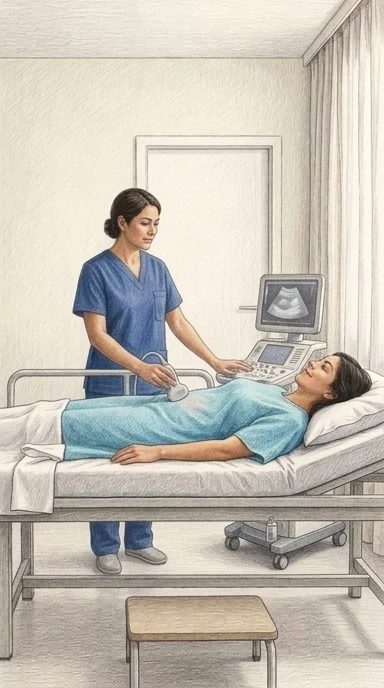
Un equipo de investigadores chinos desarrolló y probó un sistema de inteligencia artificial llamado BIRD, siglas en inglés de dispositivo de reconocimiento inteligente de mama. El sistema analiza imágenes de ecografía de mama y asigna a cada lesión una categoría de riesgo según el sistema internacional BI-RADS, el mismo que usan los radiólogos para clasificar lo que ven1. Cuando el sistema se usó como apoyo y acertó, la precisión de los radiólogos que lo consultaban mejoró de forma medible2.
Pero lo más importante no fue eso. Cuando el sistema se usó como apoyo y acertó, la precisión de los radiólogos que lo consultaban mejoró de forma medible. En un estudio prospectivo con seis radiólogos y 527 imágenes, la diferencia entre leer con y sin BIRD fue estadísticamente significativa3. En otro ensayo aleatorizado con cuatro radiólogos en formación, la precisión de un grupo pasó de 0.687 sin ayuda a 0.736 con ayuda4.
El sistema también se desplegó en la práctica real. Durante año y medio se instaló junto a los ecógrafos en 107 hospitales de China, para dar apoyo diagnóstico en tiempo real mientras se hacía el estudio5. En los 38 hospitales que enviaron datos completos, la coincidencia entre BIRD y los radiólogos locales fue de 0.702 en la escala kappa6. Esa cifra mide qué tanto coinciden, no qué tan correcto es el sistema.
Además, el equipo construyó tres modelos que intentan leer del ultrasonido algo que normalmente exige una biopsia: si la lesión es benigna o maligna, si es carcinoma invasor o carcinoma ductal in situ, y cuál es el subtipo molecular del tumor. Los dos primeros obtuvieron áreas bajo la curva de 0.965 y 0.924 respectivamente; el tercero, entre 0.753 y 0.809 según el subtipo. Son clasificadores probados contra el resultado de laboratorio, no reemplazos del laboratorio.
Hubo también una campaña de tamizaje en 10 sitios, con 6,817 mujeres. De ellas, 5,373 quedaron clasificadas como riesgo bajo, y en el grupo de riesgo alto —301 personas— se diagnosticaron tres cánceres durante un seguimiento de 13 meses7. El estudio no puede demostrar que el sistema salve vidas ni que detecte el cáncer antes que la atención habitual: solo siguió de cerca al grupo de alto riesgo, y sus autores lo describen como un estudio de viabilidad8.
Para entender por qué esto importa, hay que empezar por el problema. El cáncer de mama es el tumor más frecuente en mujeres en el mundo, y detectarlo temprano es la base para mejorar la supervivencia9. En mujeres asiáticas, que tienden a tener tejido mamario denso, la mamografía pierde sensibilidad y la ecografía se vuelve el método principal de tamizaje10. El problema es que leer una ecografía depende mucho de quién la lee: aunque el sistema BI-RADS está estandarizado, sigue siendo subjetivo, y eso lleva a biopsias innecesarias o a diagnósticos tardíos11.
La idea de que una máquina ayude a leer estos estudios no es nueva. Un repaso de diez años de investigación sobre inteligencia artificial en mamografía, cuyo resumen pudimos leer pero cuya versión completa está detrás de una suscripción, encontró que el área bajo la curva combinada de 14 estudios era 0.890, con una dispersión enorme entre unos y otros12. Esa dispersión significa que el promedio alto no se puede trasladar automáticamente a un programa nuevo: cada lugar necesita validar la herramienta con sus propias pacientes13.
Otro trabajo, también leído solo en su resumen, describe un esfuerzo parecido en mamografía: un sistema que se fue ajustando durante su uso real en un programa de seguro médico obligatorio, con más de 400,000 mamografías de 206 organizaciones médicas y 336 radiólogos participantes14. Con el tiempo y las actualizaciones, su desempeño mejoró de forma sostenida15. Los autores concluyen que la revisión continua durante el uso real es lo que hace funcionar estas herramientas16.
Un tercer estudio, igualmente leído solo en su resumen, atacó un problema muy concreto: ciertos tumores agresivos pueden parecerse en la ecografía a masas benignas comunes, y tanto los médicos como la inteligencia artificial se equivocan al distinguirlos17. Al combinar el sistema automático con la lectura del radiólogo, la precisión general mejoró18. Los propios autores advierten que la herramienta no debe reemplazar la clasificación del médico, sino acompañarla19.
El patrón que se repite en todos estos trabajos es el mismo: la inteligencia artificial funciona mejor como segundo lector o como filtro que como sustituto. Un repaso de la evidencia de una década lo dice sin rodeos: apoya la detección, no reemplaza al radiólogo20. Y las ganancias más grandes y precisas tienden a venir de estudios no aleatorizados, que es justo el patrón que produciría un sesgo21.
Así lo leemos nosotros. Lo que este estudio muestra con más fuerza no es que la máquina sea mejor que el médico, sino que puede reducir la diferencia entre un médico experimentado y uno que no lo es. En una clínica pública con poco personal especializado, eso podría significar que una lectura de ecografía de mama llegue con un nivel de riesgo ya marcado, y que el radiólogo de guardia tenga un punto de partida. Es una expectativa, no un hallazgo: el estudio midió precisión de lectura, no resultados en pacientes.
También leímos que este tipo de herramienta puede fallar de maneras que nadie nota. Si el sistema se entrenó con imágenes de ciertos hospitales y ciertas máquinas, podría comportarse distinto con pacientes de otros lugares, otros cuerpos, otros equipos. Ese detalle puede pasar desapercibido si nadie lo mide en cada centro. La forma de saber si nos equivocamos es simple: que en cada nuevo lugar se compruebe con pacientes reales y de distintos orígenes, y que los resultados se revisen con el tiempo.
Y hay algo que este estudio no resuelve: cuando una herramienta así se adopta, la paciente puede recibir un nivel de riesgo sin que nadie le explique en qué se basó. Usted puede pedir que le expliquen en palabras sencillas por qué su estudio salió con cierto nivel de riesgo, quién revisó el resultado y si existe la opción de una segunda lectura por una persona. Eso no es desconfianza: es la misma pregunta que haría con cualquier otro examen.
Nos queda una cosa por decir, y es la más útil. La próxima vez que le hagan una ecografía de mama, pregunte si el servicio cuenta con apoyo diagnóstico, si hay personal capacitado para usarlo y cómo se interpretó su estudio. Si algo no cuadra con lo que usted siente, pida otra opinión. Ninguna herramienta, por buena que sea, reemplaza esa conversación.
¿Qué le preguntaría a su médico la próxima vez?
De dónde sale cada dato de contexto, y cuánto leímos de cada documento
- Zhou, J., Si, P., Zhang, Y. et al. (2026). A non-invasive end-to-end intelligent assistance system for breast ultrasound. Nature Communications. https://doi.org/10.1038/s41467-026-73170-5 - el artículo del que trata esta nota — el artículo completo — el pasaje: "Here, we develop and test an end-to-end breast intelligent recognition device (BIRD) for women across diverse institution/population datasets."
- Zhou, J., Si, P., Zhang, Y. et al. (2026). A non-invasive end-to-end intelligent assistance system for breast ultrasound. Nature Communications. https://doi.org/10.1038/s41467-026-73170-5 - el artículo del que trata esta nota — el artículo completo — el pasaje: "The accuracy of BIRD in the internal test set is 0.837 (95% confidence interval: 0.827–0.846), which significantly improves radiologists’ accuracy in 2 reader studies ( P < 0.05)."
- Zhou, J., Si, P., Zhang, Y. et al. (2026). A non-invasive end-to-end intelligent assistance system for breast ultrasound. Nature Communications. https://doi.org/10.1038/s41467-026-73170-5 - el artículo del que trata esta nota — el artículo completo — el pasaje: "When BIRD was used for AI assistance and made correct predictions, the accuracy of the 6 radiologists was improved. Significant differences were observed in the performance of radiologists with and without AI assistance ( P = 1.02e-10) (Fig. 3b and Table 3 )."
- Zhou, J., Si, P., Zhang, Y. et al. (2026). A non-invasive end-to-end intelligent assistance system for breast ultrasound. Nature Communications. https://doi.org/10.1038/s41467-026-73170-5 - el artículo del que trata esta nota — el artículo completo — el pasaje: "In the unassisted phase, the accuracy of group A2 radiologists was 0.687 (95% CI: 0.648–0.726). With AI assistance, accuracy significantly increased to 0.736 (95% CI: 0.696–0.776, P = 0.028), accompanied by increased sensitivity from 0.508 (95% CI: 0.465–0.556) to 0.528 (95% CI: 0.469–0.623) (Fig. 3c and Table 4 )."
- Zhou, J., Si, P., Zhang, Y. et al. (2026). A non-invasive end-to-end intelligent assistance system for breast ultrasound. Nature Communications. https://doi.org/10.1038/s41467-026-73170-5 - el artículo del que trata esta nota — el artículo completo — el pasaje: "The deployment of the BIRD within the ultrasound department enabled real-time acquisition of ultrasound imagery and the delivery of simultaneous intelligent diagnostic support (Supplementary Fig. 11 and Supplementary Video 1 )."
- Zhou, J., Si, P., Zhang, Y. et al. (2026). A non-invasive end-to-end intelligent assistance system for breast ultrasound. Nature Communications. https://doi.org/10.1038/s41467-026-73170-5 - el artículo del que trata esta nota — el artículo completo — el pasaje: "BIRD is applied in breast cancer screening for 6,817 individuals and shows high consistency (Cohen’s kappa: 0.702 (95% confidence interval: 0.628-0.777)) with clinical assessments in real-world application across 107 hospitals."
- Zhou, J., Si, P., Zhang, Y. et al. (2026). A non-invasive end-to-end intelligent assistance system for breast ultrasound. Nature Communications. https://doi.org/10.1038/s41467-026-73170-5 - el artículo del que trata esta nota — el artículo completo — el pasaje: "A subsequent follow-up of the high-risk group ( n = 301), which had a median follow-up of 13 months, found that 3 patients were diagnosed with breast cancer."
- Zhou, J., Si, P., Zhang, Y. et al. (2026). A non-invasive end-to-end intelligent assistance system for breast ultrasound. Nature Communications. https://doi.org/10.1038/s41467-026-73170-5 - el artículo del que trata esta nota — el artículo completo — el pasaje: "The screening program’s verification bias and follow-up duration reflect its feasibility study design; definitive efficacy metrics will be established through future randomized controlled trials."
- Zhou, J., Si, P., Zhang, Y. et al. (2026). A non-invasive end-to-end intelligent assistance system for breast ultrasound. Nature Communications. https://doi.org/10.1038/s41467-026-73170-5 - el artículo del que trata esta nota — el artículo completo — el pasaje: "Breast cancer is the most prevalent malignancy among women worldwide 1 , 2 , with early detection being the cornerstone of improved survival."
- Zhou, J., Si, P., Zhang, Y. et al. (2026). A non-invasive end-to-end intelligent assistance system for breast ultrasound. Nature Communications. https://doi.org/10.1038/s41467-026-73170-5 - el artículo del que trata esta nota — el artículo completo — el pasaje: "In Asian women, who tend to have dense breast tissue, mammography’s sensitivity is markedly reduced, and ultrasound has emerged as the primary screening modality 3 ."
- Zhou, J., Si, P., Zhang, Y. et al. (2026). A non-invasive end-to-end intelligent assistance system for breast ultrasound. Nature Communications. https://doi.org/10.1038/s41467-026-73170-5 - el artículo del que trata esta nota — el artículo completo — el pasaje: "However, ultrasound interpretation is highly operator-dependent, and although the Breast Imaging Reporting and Data System (BI-RADS) classification is standardized, it remains subjective 4 , 5 , leading to inter-reader variability that may cause unnecessary biopsies or delayed diagnosis."
- Ciurescu S, Buciu V, Ilaș DG, Pârvănescu R, Șerban D. (2026). Ten Years of Artificial Intelligence in Screening Mammography: A Systematic Review and Meta-Analysis of Diagnostic Accuracy and Clinical Implementation (Literature Published 2015–2025). Diagnostics. 10.3390/diagnostics16183045 — solo el resumen - no se pudo obtener el texto completo — el pasaje: "Pooled standalone AUC across 14 studies and 1,214,885 examinations was 0.890 (95% CI 0.858-0.915), with I 2 = 97.4% and a 95% prediction interval of 0.731-0.960."
- Ciurescu S, Buciu V, Ilaș DG, Pârvănescu R, Șerban D. (2026). Ten Years of Artificial Intelligence in Screening Mammography: A Systematic Review and Meta-Analysis of Diagnostic Accuracy and Clinical Implementation (Literature Published 2015–2025). Diagnostics. 10.3390/diagnostics16183045 — solo el resumen - no se pudo obtener el texto completo — el pasaje: "Pooled accuracy is high on average but so dispersed that it cannot be transferred to a new programme; local validation before deployment remains necessary."
- Vasilev Y, Rumyantsev D, Vladzymyrskyy A, Omelyanskaya O, Arzamasov K, Bazhin A, et al. (2026). Implementation of an artificial intelligence-based system for mammography in the compulsory medical insurance program: results of a 3-year study. Quantitative Imaging in Medicine and Surgery. 10.21037/qims-2026-0864 — solo el resumen - no se pudo obtener el texto completo — el pasaje: "The test dataset comprised 404,502 mammograms from 206 medical organizations and three mammography equipment manufacturers. A total of 336 radiologists participated."
- Vasilev Y, Rumyantsev D, Vladzymyrskyy A, Omelyanskaya O, Arzamasov K, Bazhin A, et al. (2026). Implementation of an artificial intelligence-based system for mammography in the compulsory medical insurance program: results of a 3-year study. Quantitative Imaging in Medicine and Surgery. 10.21037/qims-2026-0864 — solo el resumen - no se pudo obtener el texto completo — el pasaje: "Over this time, AUC increased by 10.8% (from 0.83 to 0.92), accuracy by 16.9% (from 0.77 to 0.90), sensitivity by 4.8% (from 0.84 to 0.88), and specificity by 30.0% (from 0.70 to 0.91)."
- Vasilev Y, Rumyantsev D, Vladzymyrskyy A, Omelyanskaya O, Arzamasov K, Bazhin A, et al. (2026). Implementation of an artificial intelligence-based system for mammography in the compulsory medical insurance program: results of a 3-year study. Quantitative Imaging in Medicine and Surgery. 10.21037/qims-2026-0864 — solo el resumen - no se pudo obtener el texto completo — el pasaje: "Iterative testing with prospective real-world monitoring, interleaved developer updates, and radiologist feedback substantially enhanced mammography AI performance."
- Berg WA, Bandos AI, Larsen LH, Heller SL, Hooley RJ, Ha RS, et al. (2026). Training AI to Improve Distinction of Triple-Negative Invasive Breast Cancer from Cysts and Fibroadenomas on Ultrasound. Diagnostics. 10.3390/diagnostics16091354 — solo el resumen - no se pudo obtener el texto completo — el pasaje: "Circumscribed oval, hypoechoic masses are common on screening breast ultrasound (US), and the vast majority are benign. Triple-receptor negative invasive breast cancer (TNBC) can appear similar, resulting in both human and artificial intelligence (AI) interpretive errors."
- Berg WA, Bandos AI, Larsen LH, Heller SL, Hooley RJ, Ha RS, et al. (2026). Training AI to Improve Distinction of Triple-Negative Invasive Breast Cancer from Cysts and Fibroadenomas on Ultrasound. Diagnostics. 10.3390/diagnostics16091354 — solo el resumen - no se pudo obtener el texto completo — el pasaje: "Combining KDS + BIRADS significantly improved overall performance (AUC 0.98"
- Berg WA, Bandos AI, Larsen LH, Heller SL, Hooley RJ, Ha RS, et al. (2026). Training AI to Improve Distinction of Triple-Negative Invasive Breast Cancer from Cysts and Fibroadenomas on Ultrasound. Diagnostics. 10.3390/diagnostics16091354 — solo el resumen - no se pudo obtener el texto completo — el pasaje: "While KDS alone should not replace BI-RADS, when used in combination with BI-RADS, it can significantly improve specificity for highly accurate (98% sensitivity) triaging management of masses representative of those seen on screening US."
- Ciurescu S, Buciu V, Ilaș DG, Pârvănescu R, Șerban D. (2026). Ten Years of Artificial Intelligence in Screening Mammography: A Systematic Review and Meta-Analysis of Diagnostic Accuracy and Clinical Implementation (Literature Published 2015–2025). Diagnostics. 10.3390/diagnostics16183045 — solo el resumen - no se pudo obtener el texto completo — el pasaje: "A decade of evidence supports AI as a second reader and triage tool in organised screening, not as an autonomous replacement for the radiologist."
- Ciurescu S, Buciu V, Ilaș DG, Pârvănescu R, Șerban D. (2026). Ten Years of Artificial Intelligence in Screening Mammography: A Systematic Review and Meta-Analysis of Diagnostic Accuracy and Clinical Implementation (Literature Published 2015–2025). Diagnostics. 10.3390/diagnostics16183045 — solo el resumen - no se pudo obtener el texto completo — el pasaje: "The larger and more precise detection gains come from non-randomised designs, which is the pattern confounding would produce, so the randomised evidence remains the anchor."
Zhou, J., Si, P., Zhang, Y. et al. (2026). A non-invasive end-to-end intelligent assistance system for breast ultrasound. Nature Communications. https://doi.org/10.1038/s41467-026-73170-5
Quién pagó: El estudio fue financiado por el Programa Nacional Clave de Investigación y Desarrollo de China, la Comisión de Salud de la Provincia de Henan, el Programa de Ciencia y Tecnología Médica de la Comisión de Salud de la Provincia de Sichuan, el Departamento de Ciencia y Tecnología de la Provincia de Sichuan y un proyecto especial de cooperación entre la ciudad de Dazhou y escuelas; el artículo no dice si los financiadores tuvieron alguna influencia, ni menciona equipos o programas prestados por terceros.
No tome esto como consejo médico profesional.
experiment · Nature communications · the paper, 20 May 2026 · free
An AI Read Breast Scans in China. It Raised Radiologists' Accuracy.
A study in Chinese hospitals found the software classified about 84 of every 100 scans correctly. The article says it needs more testing before use outside that setting.
Short version · the longer version follows, about 7 min
Ask the weeklyAI chat
Ask me about this study: who was studied, what it found, and what it does not say.
Conversations are saved for as long as weeklyAI exists, to improve the publication. Answers come in the language you write in.
- The study at a glance
- Who
- women having breast ultrasound scans
- How many
- 6,817 individuals
- Where
- China
- When
- not stated in the passages
- Kind of study
- experiment
- Who did it
- researchers across dozens of Chinese hospitals
- The limit that matters
- The study cannot say whether this screening saved lives or caught cancer earlier than usual care.
Radiologists reading alone versus reading with BIRD's help
accuracy rose from 0.687 to 0.736
If you get a breast ultrasound, you can ask whether the clinic uses an AI second reader.
Researchers in China built a computer program called BIRD. It reads breast ultrasound images and sorts each one by BI-RADS, the standard risk scale radiologists use.
On the program's own test set, its accuracy was 0.837. That is about 84 of every 100 images sorted correctly.
In a study of 527 images, six radiologists looked at the images in two rounds. In the first round they worked alone. In the second round they had BIRD's help. One reader's accuracy rose from 0.634 to 0.877. Another went from 0.786 to 0.900. The researchers report these gains as significant.
But when BIRD got a case wrong, three of the radiologists did worse with its help. These three were one trainee, one junior and one senior radiologist. A second reader can pull you toward a mistake as easily as away from one.
The study was done in China. The article states plainly that BIRD needs more validation before clinical use outside that setting. In two international sets of images, one from Egypt and one from Spain, its accuracy fell to 0.661 and 0.604. So the 0.837 does not travel with you.
In real use, BIRD was deployed across 107 hospitals in China. In 38 hospitals with usable records, it agreed with local radiologists at a kappa of 0.702. That number describes how often the software and the doctor made the same call. It does not prove the call was right.
The researchers also ran a screening program for 6,817 people. BIRD sorted 5,373 of them as low risk and 301 as high risk. Only the high-risk group was followed, for about 13 months, and three cancers were found. The study cannot say whether this screening saved lives or caught cancer earlier than usual care.
The program also produced models that guess, from the scan alone, whether a lump is benign or malignant, and which molecular subtype a cancer is. The researchers used biopsy results as the standard to develop and check those models. They do not replace a biopsy.
If you get a breast ultrasound, you can ask whether the clinic uses an AI second reader. You can ask whether that software has been checked in your country, and on patients like you.
What this means for you
What this means for you is narrower than it sounds: the software helped radiologists read breast ultrasound images in Chinese hospitals, and it agreed with local doctors across 107 of them, but it was never checked in your country or on patients like you. If a clinic near you offers an AI second reader, ask whether it was tested here, and on whom, before you weigh its opinion.
Zhou, J., Si, P., Zhang, Y. et al. (2026). A non-invasive end-to-end intelligent assistance system for breast ultrasound. Nature Communications. https://doi.org/10.1038/s41467-026-73170-5
Who paid: The study was funded by the National Key Research and Development Program of China, the Health Commission of Henan Province, the Health Commission of Sichuan Province Medical Science and Technology Program, the Science and Technology Department of Sichuan Province, and a Dazhou City School Cooperation Special project; the article does not say whether funders had any say, and it does not mention equipment or software lent by others.
Do not take this as professional medical advice.
The longer versionThe passages copied from the paper, the pictures, and every source with how much of it we read · 1420 words · about 7 minRead it →Close
AI Helped Radiologists Read Breast Ultrasounds in a Chinese Study. Here's What It Did—and Didn't—Show.
The system raised doctors' accuracy in controlled tests and agreed with them across 107 hospitals. It has not been proven to save lives, and it is not yet validated outside China.
Breast ultrasound is hard to read. In Asian women, who tend to have dense breast tissue, mammography's sensitivity is markedly reduced, and ultrasound has emerged as the primary screening modality1. But ultrasound interpretation is highly operator-dependent, and although the Breast Imaging Reporting and Data System (BI-RADS) classification is standardized, it remains subjective, leading to inter-reader variability that may cause unnecessary biopsies or delayed diagnosis2.
A team of researchers across dozens of Chinese hospitals developed and tested an end-to-end breast intelligent recognition device (BIRD) for women across diverse institution and population datasets3. In its internal test set, BIRD's accuracy was 0.837 — meaning it agreed with the expert-assigned BI-RADS category about 84 times out of 100 — with a 95 percent confidence interval of 0.827 to 0.8464. The same paper reports that BIRD significantly improved radiologists' accuracy in two reader studies, with a P value below 0.054.
In one of those studies, when BIRD made correct predictions, the accuracy of six radiologists improved, and the difference between reading with and without AI assistance was significant at P = 1.02e-105. In a separate randomized trial, one group of trainee radiologists went from 0.687 accuracy without AI to 0.736 with it — a statistically significant gain at P = 0.028 — with sensitivity rising from 0.508 to 0.5286.
The system was then deployed for real. BIRD was applied in breast cancer screening for 6,817 individuals and showed high consistency with clinical assessments across 107 hospitals, with a Cohen's kappa of 0.7027. In the screening cohort, follow-up of the 301 people classified as high risk, over a median of 13 months, found that 3 patients were diagnosed with breast cancer8.
The limits matter as much as the numbers. Performance variations across international datasets indicate that BIRD requires further validation before clinical deployment outside the development and domestic validation setting9. The false negative rate remains within clinically acceptable thresholds for an adjunctive tool, though targeted integration of dynamic imaging context promises further refinement10. And the screening program's verification bias and follow-up duration reflect its feasibility study design; definitive efficacy metrics will be established through future randomized controlled trials11.
Here is how the technology actually works, in plain steps. The system uses one algorithm to find the lesion in the image — drawing a box around it — and a second algorithm to sort that lesion into one of the BI-RADS categories, from 1 to 5, including the subcategories 4a, 4b and 4c. It was built to run right beside the ultrasound machine, so the reading happens while the patient is still there: the deployment within the ultrasound department enabled real-time acquisition of ultrasound imagery and the delivery of simultaneous intelligent diagnostic support12. That placement is the point. A tool that speaks during the exam is different from a tool that speaks a week later.
The paper also places itself against a wider pattern in medical AI. Unlike tools cleared by the U.S. Food and Drug Administration that were evaluated solely in controlled environments, this system's prospective multi-site implementation addresses a gap where post-market surveillance data for AI devices remains scarce, offering a scalable governance template through continuous monitoring infrastructure13. In other words, the authors are arguing that the interesting part is not the accuracy number — it is that the thing was actually run in 107 hospitals and watched.
Other researchers have walked this road with mammography, not ultrasound. A review and meta-analysis covering the literature from 2015 to 2025 found that deep-learning AI for mammographic screening moved from proof of concept to randomized evaluation in a single decade (according to the summary of that study; we could read only the summary, the full paper is behind a subscription)14. That review pooled a standalone accuracy measure of 0.890 across 14 studies and 1,214,885 examinations, but the results were so dispersed that a new program could not simply inherit them — local validation before deployment remained necessary (again, from the summary only)1516. Its conclusion was pointed: a decade of evidence supports AI as a second reader and triage tool in organized screening, not as an autonomous replacement for the radiologist (summary only)17.
A separate effort in Russia took a different route: rather than testing once, it built continuous quality control into a mammography AI's implementation inside a compulsory medical insurance program, using a test set of 404,502 mammograms from 206 medical organizations and 336 radiologists (summary only)1819. Over that period the system's accuracy improved from 0.77 to 0.90 (summary only)20. Its stated limitation was that the retrospective calibration testing dataset was small and there was no external validation on independent datasets from other regions or countries (summary only)21. That is the same gap this ultrasound study names from the other direction.
A third line of work tries to sharpen AI on the hardest cases. A retrospective refinement and validation study across five academic medical centers used orthogonal ultrasound images of 1,771 breast masses 2 cm or smaller, and found that combining an AI tool with BI-RADS assessment improved overall performance, while the AI alone should not replace BI-RADS (summary only)2223. The recurring shape across all of this: AI as a second opinion, never as the only opinion.
Here is how we read it. The pattern in this family of studies is consistent — the machine is being tested as a colleague, not as a replacement, and the gains show up when a human stays in the loop. What that leads us to expect, in clinics like the ones our readers use, is a specific kind of change: not fewer doctors, but a second screen that speaks before the patient leaves the room. How would you know we are wrong? If you are offered an AI-assisted scan and the person reading it tells you they reviewed the images on their own first, and that they would have flagged the same thing without the machine — that is the good version. If instead the AI output is simply printed and signed, that is the version we are worried about. What you can do with this: when you are told a scan was read with AI assistance, ask two questions. Was the radiologist's own reading done independently, before seeing the AI result? And if you feel something that the report calls low risk, will they look again? Those two questions cost nothing and change what the machine is allowed to mean.
We also want to be honest about what we cannot yet see. The published record on AI in breast imaging is full of accuracy figures that look excellent in one setting and travel poorly to the next — this study's own numbers drop sharply on the two international datasets, and the authors say so themselves. There is a known risk in this class of system: when a confident-looking score arrives first, people can stop looking as hard, and the cases the machine misses are exactly the ones that needed the human eye. We have no evidence that happened here. But it is the thing to watch, and the thing to ask about, wherever these tools arrive.
One more thing the study did, beyond triage: it tried to read biology off the image. Three separate models were built to classify benign versus malignant lesions, invasive carcinoma versus DCIS, and five molecular subtypes of breast cancer, from ultrasound pictures alone. The paper reports these were classification models only — tested against pathology labels, not replacements for biopsy.
What this makes possible for you is narrower and more useful than a headline. It means that in some clinics, the scan you are already getting may soon come with a risk estimate attached — and that estimate is a prompt for a conversation, not a verdict. If your clinic adopts something like this, you are entitled to ask how it was tested on people like you, with the machine that will be used on you, and what happens when the machine and the radiologist disagree. The study's own authors say the definitive answers require randomized trials that have not yet been run. Until then, the honest position is the one this paper takes: promising, deployed, and still being watched.
If a scan is read with AI assistance, ask whether the radiologist formed their own reading first.
Where each piece of context comes from, and how much of it we read
- Zhou, J., Si, P., Zhang, Y. et al. (2026). A non-invasive end-to-end intelligent assistance system for breast ultrasound. Nature Communications. https://doi.org/10.1038/s41467-026-73170-5 - the article this story is about — the whole article — the passage: "In Asian women, who tend to have dense breast tissue, mammography’s sensitivity is markedly reduced, and ultrasound has emerged as the primary screening modality 3 ."
- Zhou, J., Si, P., Zhang, Y. et al. (2026). A non-invasive end-to-end intelligent assistance system for breast ultrasound. Nature Communications. https://doi.org/10.1038/s41467-026-73170-5 - the article this story is about — the whole article — the passage: "However, ultrasound interpretation is highly operator-dependent, and although the Breast Imaging Reporting and Data System (BI-RADS) classification is standardized, it remains subjective 4 , 5 , leading to inter-reader variability that may cause unnecessary biopsies or delayed diagnosis."
- Zhou, J., Si, P., Zhang, Y. et al. (2026). A non-invasive end-to-end intelligent assistance system for breast ultrasound. Nature Communications. https://doi.org/10.1038/s41467-026-73170-5 - the article this story is about — the whole article — the passage: "Here, we develop and test an end-to-end breast intelligent recognition device (BIRD) for women across diverse institution/population datasets."
- Zhou, J., Si, P., Zhang, Y. et al. (2026). A non-invasive end-to-end intelligent assistance system for breast ultrasound. Nature Communications. https://doi.org/10.1038/s41467-026-73170-5 - the article this story is about — the whole article — the passage: "The accuracy of BIRD in the internal test set is 0.837 (95% confidence interval: 0.827–0.846), which significantly improves radiologists’ accuracy in 2 reader studies ( P < 0.05)."
- Zhou, J., Si, P., Zhang, Y. et al. (2026). A non-invasive end-to-end intelligent assistance system for breast ultrasound. Nature Communications. https://doi.org/10.1038/s41467-026-73170-5 - the article this story is about — the whole article — the passage: "When BIRD was used for AI assistance and made correct predictions, the accuracy of the 6 radiologists was improved. Significant differences were observed in the performance of radiologists with and without AI assistance ( P = 1.02e-10) (Fig. 3b and Table 3 )."
- Zhou, J., Si, P., Zhang, Y. et al. (2026). A non-invasive end-to-end intelligent assistance system for breast ultrasound. Nature Communications. https://doi.org/10.1038/s41467-026-73170-5 - the article this story is about — the whole article — the passage: "In the unassisted phase, the accuracy of group A2 radiologists was 0.687 (95% CI: 0.648–0.726). With AI assistance, accuracy significantly increased to 0.736 (95% CI: 0.696–0.776, P = 0.028), accompanied by increased sensitivity from 0.508 (95% CI: 0.465–0.556) to 0.528 (95% CI: 0.469–0.623) (Fig. 3c and Table 4 )."
- Zhou, J., Si, P., Zhang, Y. et al. (2026). A non-invasive end-to-end intelligent assistance system for breast ultrasound. Nature Communications. https://doi.org/10.1038/s41467-026-73170-5 - the article this story is about — the whole article — the passage: "BIRD is applied in breast cancer screening for 6,817 individuals and shows high consistency (Cohen’s kappa: 0.702 (95% confidence interval: 0.628-0.777)) with clinical assessments in real-world application across 107 hospitals."
- Zhou, J., Si, P., Zhang, Y. et al. (2026). A non-invasive end-to-end intelligent assistance system for breast ultrasound. Nature Communications. https://doi.org/10.1038/s41467-026-73170-5 - the article this story is about — the whole article — the passage: "A subsequent follow-up of the high-risk group ( n = 301), which had a median follow-up of 13 months, found that 3 patients were diagnosed with breast cancer."
- Zhou, J., Si, P., Zhang, Y. et al. (2026). A non-invasive end-to-end intelligent assistance system for breast ultrasound. Nature Communications. https://doi.org/10.1038/s41467-026-73170-5 - the article this story is about — the whole article — the passage: "Performance variations across international datasets indicate that BIRD requires further validation before clinical deployment outside the development and domestic validation setting."
- Zhou, J., Si, P., Zhang, Y. et al. (2026). A non-invasive end-to-end intelligent assistance system for breast ultrasound. Nature Communications. https://doi.org/10.1038/s41467-026-73170-5 - the article this story is about — the whole article — the passage: "The false negative rate (FNR) remains within clinically acceptable thresholds for an adjunctive tool, though targeted integration of dynamic imaging context promises further refinement."
- Zhou, J., Si, P., Zhang, Y. et al. (2026). A non-invasive end-to-end intelligent assistance system for breast ultrasound. Nature Communications. https://doi.org/10.1038/s41467-026-73170-5 - the article this story is about — the whole article — the passage: "The screening program’s verification bias and follow-up duration reflect its feasibility study design; definitive efficacy metrics will be established through future randomized controlled trials."
- Zhou, J., Si, P., Zhang, Y. et al. (2026). A non-invasive end-to-end intelligent assistance system for breast ultrasound. Nature Communications. https://doi.org/10.1038/s41467-026-73170-5 - the article this story is about — the whole article — the passage: "The deployment of the BIRD within the ultrasound department enabled real-time acquisition of ultrasound imagery and the delivery of simultaneous intelligent diagnostic support (Supplementary Fig. 11 and Supplementary Video 1 )."
- Zhou, J., Si, P., Zhang, Y. et al. (2026). A non-invasive end-to-end intelligent assistance system for breast ultrasound. Nature Communications. https://doi.org/10.1038/s41467-026-73170-5 - the article this story is about — the whole article — the passage: "Unlike Food and Drug Administration (FDA)-cleared tools evaluated solely in controlled environments, BIRD’s prospective multi-site implementation addresses a critical regulatory gap where post-market surveillance data for AI devices remains scarce, offering a scalable governance template through continuous monitoring infrastructure."
- Ciurescu S, Buciu V, Ilaș DG, Pârvănescu R, Șerban D. (2026). Ten Years of Artificial Intelligence in Screening Mammography: A Systematic Review and Meta-Analysis of Diagnostic Accuracy and Clinical Implementation (Literature Published 2015–2025). Diagnostics. 10.3390/diagnostics16183045 — only the abstract - the full text could not be fetched — the passage: "Deep-learning artificial intelligence (AI) for mammographic screening moved from proof of concept to randomised evaluation in a single decade. We reviewed and meta-analysed its diagnostic accuracy and its effect on screening programmes, covering the literature published between 2015 and 2025."
- Ciurescu S, Buciu V, Ilaș DG, Pârvănescu R, Șerban D. (2026). Ten Years of Artificial Intelligence in Screening Mammography: A Systematic Review and Meta-Analysis of Diagnostic Accuracy and Clinical Implementation (Literature Published 2015–2025). Diagnostics. 10.3390/diagnostics16183045 — only the abstract - the full text could not be fetched — the passage: "Pooled standalone AUC across 14 studies and 1,214,885 examinations was 0.890 (95% CI 0.858-0.915), with I 2 = 97.4% and a 95% prediction interval of 0.731-0.960."
- Ciurescu S, Buciu V, Ilaș DG, Pârvănescu R, Șerban D. (2026). Ten Years of Artificial Intelligence in Screening Mammography: A Systematic Review and Meta-Analysis of Diagnostic Accuracy and Clinical Implementation (Literature Published 2015–2025). Diagnostics. 10.3390/diagnostics16183045 — only the abstract - the full text could not be fetched — the passage: "Pooled accuracy is high on average but so dispersed that it cannot be transferred to a new programme; local validation before deployment remains necessary."
- Ciurescu S, Buciu V, Ilaș DG, Pârvănescu R, Șerban D. (2026). Ten Years of Artificial Intelligence in Screening Mammography: A Systematic Review and Meta-Analysis of Diagnostic Accuracy and Clinical Implementation (Literature Published 2015–2025). Diagnostics. 10.3390/diagnostics16183045 — only the abstract - the full text could not be fetched — the passage: "A decade of evidence supports AI as a second reader and triage tool in organised screening, not as an autonomous replacement for the radiologist."
- Vasilev Y, Rumyantsev D, Vladzymyrskyy A, Omelyanskaya O, Arzamasov K, Bazhin A, et al. (2026). Implementation of an artificial intelligence-based system for mammography in the compulsory medical insurance program: results of a 3-year study. Quantitative Imaging in Medicine and Surgery. 10.21037/qims-2026-0864 — only the abstract - the full text could not be fetched — the passage: "This study aims to ensure continuous quality control of a mammography AI solution during its implementation in the CMI system by applying a novel lifecycle-based testing and monitoring methodology that addresses these specific gaps."
- Vasilev Y, Rumyantsev D, Vladzymyrskyy A, Omelyanskaya O, Arzamasov K, Bazhin A, et al. (2026). Implementation of an artificial intelligence-based system for mammography in the compulsory medical insurance program: results of a 3-year study. Quantitative Imaging in Medicine and Surgery. 10.21037/qims-2026-0864 — only the abstract - the full text could not be fetched — the passage: "The test dataset comprised 404,502 mammograms from 206 medical organizations and three mammography equipment manufacturers. A total of 336 radiologists participated."
- Vasilev Y, Rumyantsev D, Vladzymyrskyy A, Omelyanskaya O, Arzamasov K, Bazhin A, et al. (2026). Implementation of an artificial intelligence-based system for mammography in the compulsory medical insurance program: results of a 3-year study. Quantitative Imaging in Medicine and Surgery. 10.21037/qims-2026-0864 — only the abstract - the full text could not be fetched — the passage: "Over this time, AUC increased by 10.8% (from 0.83 to 0.92), accuracy by 16.9% (from 0.77 to 0.90), sensitivity by 4.8% (from 0.84 to 0.88), and specificity by 30.0% (from 0.70 to 0.91)."
- Vasilev Y, Rumyantsev D, Vladzymyrskyy A, Omelyanskaya O, Arzamasov K, Bazhin A, et al. (2026). Implementation of an artificial intelligence-based system for mammography in the compulsory medical insurance program: results of a 3-year study. Quantitative Imaging in Medicine and Surgery. 10.21037/qims-2026-0864 — only the abstract - the full text could not be fetched — the passage: "A key limitation of this study is the relatively small retrospective calibration testing dataset (100 mammograms) and the lack of external validation on independent datasets from other regions or countries."
- Berg WA, Bandos AI, Larsen LH, Heller SL, Hooley RJ, Ha RS, et al. (2026). Training AI to Improve Distinction of Triple-Negative Invasive Breast Cancer from Cysts and Fibroadenomas on Ultrasound. Diagnostics. 10.3390/diagnostics16091354 — only the abstract - the full text could not be fetched — the passage: "In an Institutional Review Board-approved HIPAA-compliant protocol, from five academic medical centers, orthogonal ultrasound images of 1771 breast masses 2 cm or smaller were acquired, consisting of cysts, complicated cysts, other benign, and malignancies."
- Berg WA, Bandos AI, Larsen LH, Heller SL, Hooley RJ, Ha RS, et al. (2026). Training AI to Improve Distinction of Triple-Negative Invasive Breast Cancer from Cysts and Fibroadenomas on Ultrasound. Diagnostics. 10.3390/diagnostics16091354 — only the abstract - the full text could not be fetched — the passage: "While KDS alone should not replace BI-RADS, when used in combination with BI-RADS, it can significantly improve specificity for highly accurate (98% sensitivity) triaging management of masses representative of those seen on screening US."
Zhou, J., Si, P., Zhang, Y. et al. (2026). A non-invasive end-to-end intelligent assistance system for breast ultrasound. Nature Communications. https://doi.org/10.1038/s41467-026-73170-5
Who paid: The study was funded by the National Key Research and Development Program of China, the Health Commission of Henan Province, the Health Commission of Sichuan Province Medical Science and Technology Program, the Science and Technology Department of Sichuan Province, and a Dazhou City School Cooperation Special project; the article does not say whether funders had any say, and it does not mention equipment or software lent by others.
Do not take this as professional medical advice.