experiment · Diagnostics (Basel, Switzerland) · la publicación, 17 abr 2026 · gratis
Una ayuda automática marcó los nódulos en las imágenes y los radiólogos dejaron escapar menos casos
El estudio es de un solo hospital de Corea del Sur y compara dos períodos, no dos grupos elegidos al azar, así que la mejora no puede atribuirse sólo al programa.
Versión breve · la versión detallada sigue, unos 6 min
Pregunte al chat de weeklyAI
Pregúnteme por este estudio: a quiénes se estudió, qué encontró y qué no dice.
Las conversaciones se guardan mientras exista weeklyAI, para mejorar la publicación. Se responde en el idioma en que usted escribe.
- El estudio, de un vistazo
- Quiénes
- Pacientes con cáncer conocido fuera del cerebro a quienes se les hizo una resonancia magnética de cerebro para buscar metástasis
- Cuántos
- 600 pacientes: 286 antes y 314 después
- Dónde
- Un hospital de Corea del Sur
- Cuándo
- Dos períodos de tres meses: enero a marzo de 2025 y mayo a julio de 2025
- Tipo de estudio
- analysis of what people did
- Quién lo hizo
- Investigadores del Hospital Universitario Nacional de Pusan, Corea del Sur
- El límite que importa
- Comparó dos períodos, no grupos sorteados; parte de la mejora pudo venir de quién leyó cada estudio
Son los porcentajes de informes que señalaron correctamente una metástasis que sí existía, en un solo hospital de Corea del Sur; no fue un ensayo con grupos sorteados, así que la mejora no puede atribuirse solo al programa.
Lo que se comparó antes y después de sumar la ayuda automática
Subió
Subió
Subió
Bajaron
Bajaron
Subió
Subió
Cuando a usted o a alguien suyo le lean una resonancia de cerebro por cáncer extendido, pregunte si están usando una ayuda automática y si esa ayuda fue probada en un estudio controlado, no sólo instalada.
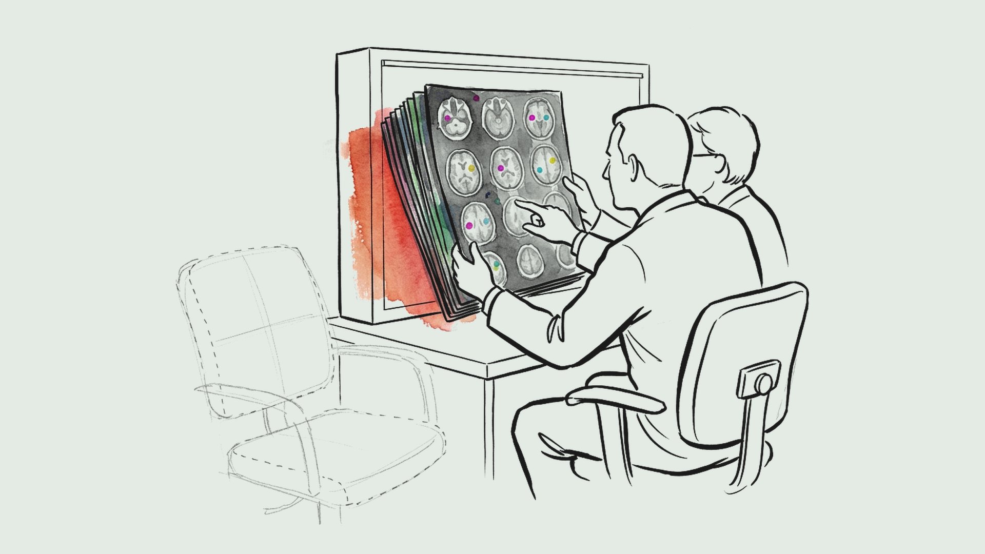
Diez radiólogos leyeron 600 resonancias magnéticas de cerebro. Eran pacientes con cáncer en otra parte del cuerpo, y el examen buscaba si el tumor se había extendido al cerebro. En 286 de esos exámenes, los radiólogos leyeron las imágenes como siempre. En los otros 314, una computadora dibujó sobre la pantalla unas marcas de color donde creía ver un nódulo.
Antes de esas marcas, los informes de los radiólogos identificaron correctamente 78 de cada 100 casos con metástasis. Después, 91 de cada 100. Los casos que se les pasaron por alto bajaron de 7 a 4 de cada 100.
También bajaron las falsas alarmas, de 12 a 6 de cada 100 exámenes. Y los casos que se les pasaron por alto bajaron de 7 a 4 de cada 100.
La diferencia fue mayor en los casos más difíciles. Cuando había un solo nódulo, la sensibilidad subió de 75 a 91 de cada 100. Cuando el nódulo más grande medía 5 milímetros o menos, la sensibilidad subió de 74 a 92 de cada 100. En cambio, cuando había varios nódulos o el más grande pasaba de 5 milímetros, los resultados fueron parecidos antes y después.
No fue un ensayo con grupos sorteados, sino una comparación de dos períodos en un mismo hospital. Distintos radiólogos leyeron distintos exámenes, y los pacientes del segundo período eran mayores. Así que parte de la mejora pudo venir de quién leyó cada estudio y no del programa.
Además, la referencia para saber quién tenía razón no fue una biopsia, sino el acuerdo de dos especialistas en imágenes, con seguimiento disponible sólo en algunos casos. Un hallazgo sutil todavía puede estar equivocado. El programa también marcó a veces lesiones benignas de vasos sanguíneos como si fueran metástasis.
Los propios radiólogos respondieron una encuesta: ocho de los diez la contestaron, y en general valoraron bien la utilidad del sistema.
Cuando a usted o a alguien suyo le lean una resonancia de cerebro por cáncer extendido, pregunte si están usando una ayuda automática y si esa ayuda fue probada en un estudio controlado, no sólo instalada.
Qué significa para usted
Cuando a usted o a alguien suyo le lean una resonancia de cerebro por cáncer extendido, pregunte si están usando una ayuda automática que marca los nódulos en la pantalla. Esa ayuda se probó sólo en un hospital de Corea del Sur, comparando dos períodos, así que la mejora observada no puede atribuirse únicamente al programa. Lo más útil sería que esa ayuda formara parte de un estudio controlado, no sólo instalada.
Roh, J., Baek, H. J., Baik, S. K. et al. (2026). Report-Level Impact of DL Assistance on Teleradiology Quality Support for Brain Metastases: Real-World Clinical Practice at a Single Tertiary Center. Diagnostics. https://doi.org/10.3390/diagnostics16081211
Quién pagó: El estudio fue financiado por una subvención de investigación de 2026 del Hospital Universitario Nacional de Pusan; el artículo declara no tener conflictos de interés y no indica que ningún financiador o proveedor haya intervenido en el diseño, el análisis o la redacción.
No tome esto como consejo médico profesional.
Versión detalladaLos pasajes copiados del artículo, las ilustraciones y cada fuente con cuánto leímos de ella · 1276 palabras · unos 6 minLeerla →Cerrar
La ayuda automática en la lectura de escaneos cerebrales: lo que mostró un hospital de Corea del Sur
Un estudio comparó informes de telerradiología antes y después de sumar un programa de detección automática de metástasis cerebrales. Los errores bajaron en el papel, pero el diseño no permite decir que el programa los causó.
En pacientes con cáncer conocido fuera del cerebro que se hicieron un escaneo cerebral para buscar metástasis, un programa de detección automática se sumó a la lectura de las imágenes en un servicio de telerradiología subcontratado. El estudio comparó dos períodos: 286 personas cuyos escaneos se leyeron sin la ayuda del programa y 314 cuyos escaneos se leyeron con ella. En el segundo período, los informes identificaron más metástasis que realmente existían y también descartaron mejor las que no existían. La proporción de informes que señalaron correctamente una metástasis presente subió de 77.7% a 90.8%; la de informes que descartaron correctamente una metástasis ausente, de 82.3% a 90.8%; y la de informes acertados en general, de 80.8% a 90.8%1. Los errores por exceso bajaron de 11.9% a 5.7% y los errores por omisión, de 7.3% a 3.5%2.
La mejora fue más notoria justo donde el ojo suele fallar. En los casos con una sola metástasis, la detección pasó de 74.6% a 91.2%; en los casos con la lesión más grande de 5 milímetros o menos, de 74.3% a 92.0%. En cambio, cuando había varias metástasis o la lesión mayor superaba los 5 milímetros, la detección fue parecida en ambos períodos3. Ocho de los diez radiólogos del servicio contestaron una encuesta: la utilidad percibida y el apoyo al diagnóstico promediaron más de 4 puntos sobre 5, incluida la ayuda para detectar lesiones y la facilidad de la interfaz4.
Conviene detenerse en lo que este estudio no puede decir. No fue un ensayo aleatorizado: comparó dos grupos de personas distintas, en dos momentos distintos, sin sortear quién recibía qué. Los propios autores advierten que no se puede descartar que otros factores —las características de los pacientes, la mezcla de casos, el paso del tiempo o quién leyó cada estudio— expliquen parte de la diferencia5. Tampoco se modeló el hecho de que cada escaneo lo leyó un médico distinto, lo que pudo influir en los resultados6. Y el programa a veces marcó como metástasis lesiones vasculares benignas7, mientras que las lesiones pequeñas o solitarias con realce tenue siguieron escapándose8. El estudio midió informes, no si los pacientes vivieron más o mejor.
Para entender de qué se trata: la resonancia magnética con contraste es la prueba de referencia para detectar metástasis cerebrales, y cada vez se combinan dos tipos de secuencia —una que resalta la sangre blanca y otra que la suprime— con programas de aprendizaje automático que buscan y delimitan lesiones solas, porque esas imágenes las hacen más visibles9. El programa usado aquí es un sistema comercial basado en una arquitectura llamada nnU-Net en 3D, diseñado para detectar metástasis en esas dos secuencias10. En el período con ayuda, las marcas de detección aparecían solas en la pantalla del visor para cada estudio, y cada radiólogo podía tomarlas en cuenta o ignorarlas para su informe final11.
Esto importa porque las metástasis cerebrales son frecuentes en personas con cáncer diseminado y pesan mucho en el pronóstico y en las decisiones de tratamiento; detectarlas y medirlas bien es parte de elegir la estrategia para cada paciente12. En un servicio subcontratado, donde cada estudio lo lee un médico distinto y las formaciones son muy diversas, esa tarea es especialmente frágil. El artículo recoge otros trabajos que ya habían mostrado resultados prometedores, pero en condiciones más controladas: describe un modelo entrenado y validado en varias instituciones que detectó metástasis con alta sensibilidad13, y un estudio multicéntrico con asignación al azar que reportó que la asistencia automática mejoraba la precisión y la eficiencia de los radiólogos al delimitar metástasis, en un entorno académico14. Lo nuevo aquí no es el programa, sino haberlo medido en el flujo real de un servicio tercerizado, con informes de texto libre.
Vale la pena mirar cómo se comportan otros usos de la inteligencia artificial en medicina, porque el patrón se repite. Un sistema aprobado para el diagnóstico de enfermedad pulmonar intersticial, usado en 209 pacientes de 47 centros estadounidenses, se asoció con más pacientes que recibieron tratamiento específico y con procedimientos invasivos que se evitaron, según el resumen de ese trabajo; pudimos leer solo el resumen151617. En cáncer de cuello uterino, un sistema que analiza imágenes de laboratorio identificó un subtipo agresivo y poco diagnosticado con alta sensibilidad en una validación grande, también según el resumen18. Y en Inglaterra, análisis de sangre con modelos de aprendizaje automático priorizaron a los pacientes de mayor riesgo en las vías de sospecha de cáncer, según el resumen de ese estudio19. En todos los casos, el patrón es el mismo: la herramienta ordena, prioriza o señala; la decisión sigue siendo humana.
Así lo leemos nosotros. Cuando una tarea se repite cientos de veces y cada caso lo ve un médico distinto, la atención tiende a repartirse de manera desigual: los casos del medio del recorrido se vuelven rutina, y lo pequeño y aislado —lo que exige detenerse— es lo que más se escapa. Por eso nos parece significativo que la mejora apareciera justo en las lesiones solitarias y pequeñas, y no en las múltiples o grandes. Si eso se sostiene, esperaríamos que en servicios con alta carga de trabajo y sin control de quién lee qué, la ayuda automática funcione sobre todo como una red que obliga a mirar dos veces lo que el ojo ya daba por visto. Nos equivocaríamos si en otros servicios la mejora resultara igual de grande en los casos con muchas metástasis o con lesiones grandes, o si apareciera también en médicos con poca carga de trabajo. Lo que usted puede hacer con esto: cuando le entreguen el informe de un escaneo cerebral, pregunte si se revisó con ayuda automática y si se buscaron específicamente las lesiones más pequeñas; si no se hizo, pedir una segunda lectura es razonable.
Hay una segunda lectura que nos parece útil. En un servicio subcontratado, con médicos de formación muy distinta y sin auditoría sistemática, existe el riesgo de que la herramienta termine funcionando como un sello: el informe lleva la marca de que hubo ayuda, aunque nadie verifique caso por caso si la alerta era correcta. Nos equivocaríamos si los informes documentaran explícitamente cómo se usó la ayuda, si existieran auditorías independientes de los casos discordantes, o si los médicos con menos experiencia no mostraran más dependencia de la alerta que los expertos. Usted puede preguntar quién responde si la ayuda automática se equivoca, y pedir que quede escrito en el informe si la alerta fue revisada o solo aceptada.
También vale preguntar si su estudio fue leído a distancia, si se usó ayuda automática y quién firmó el informe. Esa información puede quedar en su expediente, y no cuesta nada pedirla.
Los autores ya señalan el paso siguiente: hacen falta estudios prospectivos y por lesión individual, que aclaren cómo integrar estas herramientas con la formación de los médicos, la organización del trabajo y la auditoría estructurada20. El programa usado es comercial y está en revisión regulatoria, así que sus pesos y su código no son públicos; aun así, su configuración y su forma de entrenamiento se pueden reproducir a partir de publicaciones citadas y componentes abiertos.
La próxima vez que alguien de su familia se haga un escaneo cerebral por sospecha de metástasis, usted ya tiene una pregunta concreta que hacer: ¿este informe se revisó con ayuda automática, y se buscaron a propósito las lesiones más pequeñas?
De dónde sale cada dato de contexto, y cuánto leímos de cada documento
- Roh, J., Baek, H. J., Baik, S. K. et al. (2026). Report-Level Impact of DL Assistance on Teleradiology Quality Support for Brain Metastases: Real-World Clinical Practice at a Single Tertiary Center. Diagnostics. https://doi.org/10.3390/diagnostics16081211 - el artículo del que trata esta nota — el artículo completo — el pasaje: "Compared with the pre-DL group, the post-DL group showed higher sensitivity (77.7% vs. 90.8%, p < 0.001), specificity (82.3% vs. 90.8%, p = 0.002), accuracy (80.8% vs. 90.8%, p < 0.001), positive predictive value (68.2% vs. 85.7%, p < 0.001), and negative predictive value (88.3% vs. 94.2%, p = 0.011)."
- Roh, J., Baek, H. J., Baik, S. K. et al. (2026). Report-Level Impact of DL Assistance on Teleradiology Quality Support for Brain Metastases: Real-World Clinical Practice at a Single Tertiary Center. Diagnostics. https://doi.org/10.3390/diagnostics16081211 - el artículo del que trata esta nota — el artículo completo — el pasaje: "False-positive and false-negative rates were lower after DL implementation (11.9% vs. 5.7%, p = 0.009; 7.3% vs. 3.5%, p = 0.045)."
- Roh, J., Baek, H. J., Baik, S. K. et al. (2026). Report-Level Impact of DL Assistance on Teleradiology Quality Support for Brain Metastases: Real-World Clinical Practice at a Single Tertiary Center. Diagnostics. https://doi.org/10.3390/diagnostics16081211 - el artículo del que trata esta nota — el artículo completo — el pasaje: "Sensitivity gains were most pronounced for cases with single metastasis (74.6% vs. 91.2%, p = 0.007) and with the largest lesion ≤ 5 mm (74.3% vs. 92.0%, p = 0.004), whereas sensitivity was similar for multiple metastases and for cases with a largest lesion > 5 mm."
- Roh, J., Baek, H. J., Baik, S. K. et al. (2026). Report-Level Impact of DL Assistance on Teleradiology Quality Support for Brain Metastases: Real-World Clinical Practice at a Single Tertiary Center. Diagnostics. https://doi.org/10.3390/diagnostics16081211 - el artículo del que trata esta nota — el artículo completo — el pasaje: "Overall, mean item scores were above 4.0 on a 5-point Likert scale for perceived usability and diagnostic support ( Table 4 ), including lesion detection assistance (mean, 4.88) and interface usability (mean, 5.00)."
- Roh, J., Baek, H. J., Baik, S. K. et al. (2026). Report-Level Impact of DL Assistance on Teleradiology Quality Support for Brain Metastases: Real-World Clinical Practice at a Single Tertiary Center. Diagnostics. https://doi.org/10.3390/diagnostics16081211 - el artículo del que trata esta nota — el artículo completo — el pasaje: "Further, as a retrospective comparison of two independent, non-paired cohorts without randomization, residual confounding related to patient characteristics, case mix, temporal effects, and reader-level factors cannot be excluded."
- Roh, J., Baek, H. J., Baik, S. K. et al. (2026). Report-Level Impact of DL Assistance on Teleradiology Quality Support for Brain Metastases: Real-World Clinical Practice at a Single Tertiary Center. Diagnostics. https://doi.org/10.3390/diagnostics16081211 - el artículo del que trata esta nota — el artículo completo — el pasaje: "Because the study was designed as a pragmatic workflow evaluation rather than a multi-reader multi-case experiment, reader-level clustering was not explicitly modeled."
- Roh, J., Baek, H. J., Baik, S. K. et al. (2026). Report-Level Impact of DL Assistance on Teleradiology Quality Support for Brain Metastases: Real-World Clinical Practice at a Single Tertiary Center. Diagnostics. https://doi.org/10.3390/diagnostics16081211 - el artículo del que trata esta nota — el artículo completo — el pasaje: "The DL system occasionally flagged benign low-flow vascular lesions (e.g., developmental venous anomaly and capillary telangiectasia) as metastases, as reflected in the McNemar test results ( Figure 6 and Figure 7 )."
- Roh, J., Baek, H. J., Baik, S. K. et al. (2026). Report-Level Impact of DL Assistance on Teleradiology Quality Support for Brain Metastases: Real-World Clinical Practice at a Single Tertiary Center. Diagnostics. https://doi.org/10.3390/diagnostics16081211 - el artículo del que trata esta nota — el artículo completo — el pasaje: "Despite these differences, false negatives persisted for small (≤5 mm) and/or solitary lesions with faint enhancement ( Figure 3 , Figure 4 and Figure 5 )."
- Roh, J., Baek, H. J., Baik, S. K. et al. (2026). Report-Level Impact of DL Assistance on Teleradiology Quality Support for Brain Metastases: Real-World Clinical Practice at a Single Tertiary Center. Diagnostics. https://doi.org/10.3390/diagnostics16081211 - el artículo del que trata esta nota — el artículo completo — el pasaje: "Contrast-enhanced magnetic resonance imaging (MRI) is the gold standard for detecting brain metastases. However, dual-sequence protocols combining white (e.g., MPRAGE) and black (e.g., SPACE/CUBE) blood imaging are increasingly being integrated with deep learning (DL) algorithms for automated lesion detection and segmentation because they enhance lesion conspicuity [ 3 , 4 , 5 , 6 ]."
- Roh, J., Baek, H. J., Baik, S. K. et al. (2026). Report-Level Impact of DL Assistance on Teleradiology Quality Support for Brain Metastases: Real-World Clinical Practice at a Single Tertiary Center. Diagnostics. https://doi.org/10.3390/diagnostics16081211 - el artículo del que trata esta nota — el artículo completo — el pasaje: "The DL-based detection system utilized a commercially developed 3D nnU-Net-based DL architecture designed to detect brain metastases on contrast-enhanced dual-sequence MRI (3D gradient-echo and 3D turbo spin-echo with black-blood technique)."
- Roh, J., Baek, H. J., Baik, S. K. et al. (2026). Report-Level Impact of DL Assistance on Teleradiology Quality Support for Brain Metastases: Real-World Clinical Practice at a Single Tertiary Center. Diagnostics. https://doi.org/10.3390/diagnostics16081211 - el artículo del que trata esta nota — el artículo completo — el pasaje: "In the post-DL period, DL-generated detection overlays were automatically displayed within the PACS viewer for every examination, and radiologists could incorporate or disregard the overlays at their discretion for the final interpretation."
- Roh, J., Baek, H. J., Baik, S. K. et al. (2026). Report-Level Impact of DL Assistance on Teleradiology Quality Support for Brain Metastases: Real-World Clinical Practice at a Single Tertiary Center. Diagnostics. https://doi.org/10.3390/diagnostics16081211 - el artículo del que trata esta nota — el artículo completo — el pasaje: "Brain metastases are common in patients with systemic cancer and critically influence their prognosis and therapeutic planning [ 1 ]. Accurate detection, quantification, and longitudinal monitoring of these lesions are essential to determine individualized treatment strategies [ 2 ]."
- Roh, J., Baek, H. J., Baik, S. K. et al. (2026). Report-Level Impact of DL Assistance on Teleradiology Quality Support for Brain Metastases: Real-World Clinical Practice at a Single Tertiary Center. Diagnostics. https://doi.org/10.3390/diagnostics16081211 - el artículo del que trata esta nota — el artículo completo — el pasaje: "A data-centric DL model trained and validated by Topff et al. across multiple institutions demonstrated high sensitivity and generalizability for detecting brain metastases on contrast-enhanced MR images [ 7 ]."
- Roh, J., Baek, H. J., Baik, S. K. et al. (2026). Report-Level Impact of DL Assistance on Teleradiology Quality Support for Brain Metastases: Real-World Clinical Practice at a Single Tertiary Center. Diagnostics. https://doi.org/10.3390/diagnostics16081211 - el artículo del que trata esta nota — el artículo completo — el pasaje: "Similarly, Luo et al. conducted a multi-center randomized crossover study and reported the benefits of artificial intelligence (AI) assistance in improving the accuracy and efficiency of brain metastasis segmentation by radiologists in controlled academic settings [ 8 ]."
- Tambe AP, Boente RD, George G, Kheir F, Tahamtani Omran O, Selvan KC. (2026). Clinical Utility of an FDA-Authorized Artificial Intelligence Imaging Platform in Interstitial Lung Disease Diagnosis. Diagnostics. 10.3390/diagnostics16152445 — solo el resumen - no se pudo obtener el texto completo — el pasaje: "There were 209 patients that underwent evaluation with Fibresolve across 47 medical centers. Of those, 16 were academic centers ( n = 57 patients), and 31 were community-based ( n = 152 patients)."
- Tambe AP, Boente RD, George G, Kheir F, Tahamtani Omran O, Selvan KC. (2026). Clinical Utility of an FDA-Authorized Artificial Intelligence Imaging Platform in Interstitial Lung Disease Diagnosis. Diagnostics. 10.3390/diagnostics16152445 — solo el resumen - no se pudo obtener el texto completo — el pasaje: "The percentage of patients receiving ILD-specific pharmacotherapy increased significantly following Fibresolve utilization (12.5% to 44.4%, p"
- Tambe AP, Boente RD, George G, Kheir F, Tahamtani Omran O, Selvan KC. (2026). Clinical Utility of an FDA-Authorized Artificial Intelligence Imaging Platform in Interstitial Lung Disease Diagnosis. Diagnostics. 10.3390/diagnostics16152445 — solo el resumen - no se pudo obtener el texto completo — el pasaje: "there was a statistically significant increase in the percentage of patients receiving guideline-directed therapy for ILD, and previously-planned invasive diagnostic procedures were avoided in many cases."
- Yang J, He Q, Peng J, Wang Y, Li J, Li H, et al. (2026). Artificial intelligence‐assisted histopathological diagnosis of endocervical gastric‐type adenocarcinoma: a multicenter model development and validation study. The Journal of Pathology: Clinical Research. 10.1002/2056-4538.70113 — solo el resumen - no se pudo obtener el texto completo — el pasaje: "In large-scale real-world deployment (Stage III, n = 7,056), GASPath achieved a balanced accuracy of 0.953, with 100% sensitivity for GAS (45/45 cases correctly identified)."
- Neal M, Dean M, Duffy S, Ferguson RE, Horan P, Johnston C, et al. (2026). Real-World Validation of PinPoint Blood Tests in the NHS: Multivariable Machine Learning to Predict Cancer Risk in Primary Care Urgent Referrals. Mayo Clinic Proceedings: Digital Health. 10.1016/j.mcpdig.2026.100382 — solo el resumen - no se pudo obtener el texto completo — el pasaje: "Prioritization of the 10% of highest-risk patients would reduce the number needed to investigate to detect one cancer by a factor of 2.6-6.1."
- Roh, J., Baek, H. J., Baik, S. K. et al. (2026). Report-Level Impact of DL Assistance on Teleradiology Quality Support for Brain Metastases: Real-World Clinical Practice at a Single Tertiary Center. Diagnostics. https://doi.org/10.3390/diagnostics16081211 - el artículo del que trata esta nota — el artículo completo — el pasaje: "Future prospective and lesion-level studies should clarify how such tools can be integrated with reader training, workflow alignment, and structured auditing strategies."
Roh, J., Baek, H. J., Baik, S. K. et al. (2026). Report-Level Impact of DL Assistance on Teleradiology Quality Support for Brain Metastases: Real-World Clinical Practice at a Single Tertiary Center. Diagnostics. https://doi.org/10.3390/diagnostics16081211
Quién pagó: El estudio fue financiado por una subvención de investigación de 2026 del Hospital Universitario Nacional de Pusan; el artículo declara no tener conflictos de interés y no indica que ningún financiador o proveedor haya intervenido en el diseño, el análisis o la redacción.
No tome esto como consejo médico profesional.
Los hallazgos de otros estudios que aquí se mencionan los conocemos por este documento, que fue el que leímos; no abrimos cada uno de esos estudios.
experiment · Diagnostics (Basel, Switzerland) · the paper, 17 Apr 2026 · free
Scans Read With AI Help Caught More Cancer Spread
In one hospital's before-and-after comparison, scans read with the aid caught more cancer spread and raised fewer false alarms. The study cannot prove the aid caused it.
Short version · the longer version follows, about 7 min
Ask the weeklyAI chat
Ask me about this study: who was studied, what it found, and what it does not say.
Conversations are saved for as long as weeklyAI exists, to improve the publication. Answers come in the language you write in.
- The study at a glance
- Who
- people with cancer who had brain MRI scans read by remote doctors
- How many
- 600 patients
- Where
- Pusan National University Yangsan Hospital, Republic of Korea
- When
- two three-month periods in 2025
- Kind of study
- analysis of what people did
- Who did it
- Pusan National University Yangsan Hospital
- The limit that matters
- Two separate groups, not randomized, so it cannot prove the aid caused the improvement.
These are the shares of scans with real cancer spread that the doctors' reports caught, before and after the computer aid was added; the study compared two separate groups of patients, not the same doctors reading the same scans twice.
Reports before the computer aid vs. reports after it
Caught more real cancer spread (77.7% vs. 90.8%)
Fewer false alarms (11.9% vs. 5.7%)
Fewer missed cases (7.3% vs. 3.5%)
The gain was big for single spots (74.6% vs. 91.2%) and small for many spots
The gain was big for the smallest spots (74.3% vs. 92.0%) and small for larger ones
When you or someone you love has a brain scan for cancer spread, ask whether a computer aid is being used, and whether it has been tested in a controlled study.
Before a computer aid was added to their screens, doctors reading a brain scan caught cancer that had spread there about 78 times out of 100 scans that truly had spots. After the aid was turned on, they caught about 91 out of 100.
Ten teleradiologists read 600 brain MRI scans for cancer spread at one hospital in the Republic of Korea. These are doctors who read scans remotely for the hospital. They read 286 scans before the aid and 314 after, over two three-month periods in 2025.
The aid marked spots on the images with colored overlays. Doctors could use the marks or ignore them, and they kept full responsibility for the report.
The false alarms fell too. Before the aid, about 12 of every 100 scans were called positive when there was no spread. After, about 6 of every 100. Missed cases fell from about 7 to about 4 of every 100.
The biggest gains came on the hardest scans. For single spots, the doctors caught about 75 of every 100 before and about 91 after. For spots 5 millimeters or smaller, they caught about 74 before and about 92 after.
But this was one hospital's comparison of two periods, not a randomized trial, so it cannot prove the aid caused the improvement. Different doctors read different scans, and the study did not account for that, so some of the difference may come from who happened to be reading.
The aid also sometimes flagged harmless blood vessel lesions as cancer. And the study did not measure whether patients lived longer or changed treatment; it measured only what the reports said.
The standard used to judge the reports was agreement between expert radiologists, not biopsy. Follow-up scans were reviewed for only some unclear cases. So a subtle finding can still be wrong.
When you or someone you love has a brain scan for cancer spread, ask whether a computer aid is being used, and whether it has been tested in a controlled study.
What this means for you
One hospital in the Republic of Korea tried this, and its own report says the improvement may owe something to which doctor happened to read which scan. So when a scan is done for cancer that may have spread to the brain, you can ask whether a computer aid is on the screen, and whether anyone has tested it where you are treated. A professional is the one to ask what it means for the report in front of you.
Roh, J., Baek, H. J., Baik, S. K. et al. (2026). Report-Level Impact of DL Assistance on Teleradiology Quality Support for Brain Metastases: Real-World Clinical Practice at a Single Tertiary Center. Diagnostics. https://doi.org/10.3390/diagnostics16081211
Who paid: The study was supported by a 2026 research grant from Pusan National University Yangsan Hospital; the article declares no conflicts of interest and does not state that any funder or vendor had a role in the design, analysis, or reporting.
Do not take this as professional medical advice.
The longer versionThe passages copied from the paper, the pictures, and every source with how much of it we read · 1305 words · about 7 minRead it →Close
AI Spotting Small Brain Tumors: One Hospital's Before-and-After
In one hospital's teleradiology service, reports missed fewer small spots during a period when a computer marked them first — but the doctors still decided.
Brain metastases are common in people whose cancer has spread, and finding them accurately shapes what treatment a person is offered. At Pusan National University Yangsan Hospital in the Republic of Korea, researchers compared two three-month periods of brain MRI reports read by outside teleradiologists: 286 patients before a deep-learning detection tool was added to the reading workflow, and 314 patients after. In the second period, report-level sensitivity rose from 77.7% to 90.8%, and false-positive reports fell from 11.9% to 5.7%12. The gains were concentrated where a reader is most likely to skim past something: single metastases rose from 74.6% to 91.2%, and lesions 5 mm or smaller from 74.3% to 92.0%, while cases with many spots or a large obvious lesion barely changed3. The study was supported by a 2026 research grant from the hospital, and the authors declare no conflicts of interest.
Contrast-enhanced MRI is the standard way to look for brain metastases, and the hospital used a dual-sequence protocol that pairs a white-blood image with a black-blood one, which makes small lesions stand out against blood vessels4. The software was a commercially developed 3D nnU-Net system trained on scans from other institutions, and in the second period it automatically drew colored markers over suspected spots in the viewing software for every scan; radiologists could accept or ignore each mark56. Ten board-certified teleradiologists read the scans — most with between two and twenty-seven years of experience, and only two fellowship-trained in neuroradiology — and each scan was read by one of them, assigned by the vendor's routine schedule, not by the researchers7. Two faculty neuroradiologists, blinded to both the teleradiology reports and the software's output, built the reference standard by consensus, with a senior neuroradiologist as adjudicator when they could not agree.
This is not the first attempt of its kind. The article describes a study in which a model trained and validated across multiple institutions by Topff and colleagues showed high sensitivity and generalizability for detecting brain metastases on contrast-enhanced MR images8. The article describes a study in which Luo and colleagues ran a multi-center randomized crossover study and reported that AI assistance improved the accuracy and efficiency of brain metastasis segmentation by radiologists in controlled academic settings9. The difference here is the setting: routine, outsourced, free-text reporting, where nobody was comparing each radiologist against themselves. The authors say future prospective and lesion-level studies should clarify how such tools fit with reader training, workflow alignment, and structured auditing10. The radiologists who answered a post-reading survey rated the tool's usability and diagnostic support above 4.0 on a 5-point scale, including lesion detection assistance at a mean of 4.88 and interface usability at 5.00, though the authors call these findings descriptive and exploratory because only eight of ten responded1112.
The study's own limits matter as much as its numbers. It was a retrospective comparison of two separate groups, not a randomized trial, so leftover differences from patient mix, timing, and who happened to read which scan cannot be ruled out13. Reader-level clustering was not modeled, meaning some of the gap may come from which doctor read which scan rather than from the tool7. The reference standard was expert consensus, not biopsy, and follow-up imaging was available only for some ambiguous cases. The reports were free text, so the researchers could not check performance lesion by lesion. And the tool made its own mistakes: it sometimes flagged harmless low-flow vascular tangles as tumors, and it still missed small, faint, solitary spots1415.
Here is how we read it. When a machine's marks are placed in front of a trained professional, the professional still decides what to accept, change, or wave away — and the machine's own errors are the ones most likely to survive into the report, because a confident-looking marker is hard to argue with. We expect the teleradiologists here did not treat the overlays as neutral suggestions. Some probably deferred to a mark even when their own reading of a faint spot disagreed, and the benign vessel tangles flagged as tumors likely reached some reports unchallenged. What would show we are wrong: reports from the second period showing radiologists overruling the software at a meaningful rate, or evidence that flagged false marks were routinely caught and corrected before the report was written. What you can do with that: if a scan report mentions a computer-assisted reading, it is fair to ask whether the radiologist reviewed the marked areas themselves and whether any flagged spot was confirmed on a second sequence or a follow-up scan. Asking for a second opinion on a borderline finding is a normal request, not an accusation.
There is a second pattern worth naming. Tools built to catch what tired or rushed people miss tend to work best on the plain, easily-skimmed case, while the genuinely ambiguous case still rests on the person's judgment — and the tool's own mistakes cluster exactly where judgment is hardest. That is what the numbers here look like: the improvement landed on single small spots, not on scans full of obvious lesions, and the software's errors landed on look-alike benign findings. So when a report says a single small spot was found or ruled out, that is the situation where the aid mattered most — worth asking whether a follow-up scan in a few months is planned. When a report mentions an uncertain or borderline finding, ask what else it could be besides a tumor.
One more thing, about the people doing the reading. People tend to accept a machine's help more readily than a machine's authority, and they want a person they can reach when something goes wrong — a number cannot be argued with or held to account. The radiologists here rated the tool highly for support, but the survey items about leaning on it long-term scored lower and varied more; and the authors themselves write that radiologist judgment remains essential for subtle or borderline findings. So ask who read your scan and whether that person can be reached with questions. A tool that flags spots is not a substitute for a named clinician who will discuss what the finding means for treatment.
One practical note for anyone trying to read the original: the software's trained weights and source code are not public because the model is proprietary and under regulatory review, so independent replication is limited. The authors say de-identified imaging data and summary performance figures are available from the corresponding author on reasonable request, subject to institutional approval.
What this study did not measure is as important as what it did. It did not look at whether patients lived longer, changed treatment, or did better in any way; it measured what the reports said. It did not test the tool anywhere outside this one hospital in the Republic of Korea, and not against any other vendor's service.
Where does that leave you, in a clinic in Latin America or a hospital in Canada or the United States? The honest answer is that this is one hospital's experience, and no one can tell you your clinic's reading service works the same way. But it gives you a question you did not have before: when a scan is read outside your hospital, ask whether a computer aid is used and whether the person reading it reviews the marked areas themselves. If your report flags a single small spot, ask what follow-up is planned. If it describes something uncertain, ask what else it could be. The technology may be coming to your clinic whether or not anyone asks — you can be the person who asks how it is being checked.
Where each piece of context comes from, and how much of it we read
- Roh, J., Baek, H. J., Baik, S. K. et al. (2026). Report-Level Impact of DL Assistance on Teleradiology Quality Support for Brain Metastases: Real-World Clinical Practice at a Single Tertiary Center. Diagnostics. https://doi.org/10.3390/diagnostics16081211 - the article this story is about — the whole article — the passage: "Compared with the pre-DL group, the post-DL group showed higher sensitivity (77.7% vs. 90.8%, p < 0.001), specificity (82.3% vs. 90.8%, p = 0.002), accuracy (80.8% vs. 90.8%, p < 0.001), positive predictive value (68.2% vs. 85.7%, p < 0.001), and negative predictive value (88.3% vs. 94.2%, p = 0.011)."
- Roh, J., Baek, H. J., Baik, S. K. et al. (2026). Report-Level Impact of DL Assistance on Teleradiology Quality Support for Brain Metastases: Real-World Clinical Practice at a Single Tertiary Center. Diagnostics. https://doi.org/10.3390/diagnostics16081211 - the article this story is about — the whole article — the passage: "False-positive and false-negative rates were lower after DL implementation (11.9% vs. 5.7%, p = 0.009; 7.3% vs. 3.5%, p = 0.045)."
- Roh, J., Baek, H. J., Baik, S. K. et al. (2026). Report-Level Impact of DL Assistance on Teleradiology Quality Support for Brain Metastases: Real-World Clinical Practice at a Single Tertiary Center. Diagnostics. https://doi.org/10.3390/diagnostics16081211 - the article this story is about — the whole article — the passage: "Sensitivity gains were most pronounced for cases with single metastasis (74.6% vs. 91.2%, p = 0.007) and with the largest lesion ≤ 5 mm (74.3% vs. 92.0%, p = 0.004), whereas sensitivity was similar for multiple metastases and for cases with a largest lesion > 5 mm."
- Roh, J., Baek, H. J., Baik, S. K. et al. (2026). Report-Level Impact of DL Assistance on Teleradiology Quality Support for Brain Metastases: Real-World Clinical Practice at a Single Tertiary Center. Diagnostics. https://doi.org/10.3390/diagnostics16081211 - the article this story is about — the whole article — the passage: "Contrast-enhanced magnetic resonance imaging (MRI) is the gold standard for detecting brain metastases. However, dual-sequence protocols combining white (e.g., MPRAGE) and black (e.g., SPACE/CUBE) blood imaging are increasingly being integrated with deep learning (DL) algorithms for automated lesion detection and segmentation because they enhance lesion conspicuity [ 3 , 4 , 5 , 6 ]."
- Roh, J., Baek, H. J., Baik, S. K. et al. (2026). Report-Level Impact of DL Assistance on Teleradiology Quality Support for Brain Metastases: Real-World Clinical Practice at a Single Tertiary Center. Diagnostics. https://doi.org/10.3390/diagnostics16081211 - the article this story is about — the whole article — the passage: "The DL-based detection system utilized a commercially developed 3D nnU-Net-based DL architecture designed to detect brain metastases on contrast-enhanced dual-sequence MRI (3D gradient-echo and 3D turbo spin-echo with black-blood technique)."
- Roh, J., Baek, H. J., Baik, S. K. et al. (2026). Report-Level Impact of DL Assistance on Teleradiology Quality Support for Brain Metastases: Real-World Clinical Practice at a Single Tertiary Center. Diagnostics. https://doi.org/10.3390/diagnostics16081211 - the article this story is about — the whole article — the passage: "In the post-DL period, DL-generated detection overlays were automatically displayed within the PACS viewer for every examination, and radiologists could incorporate or disregard the overlays at their discretion for the final interpretation."
- Roh, J., Baek, H. J., Baik, S. K. et al. (2026). Report-Level Impact of DL Assistance on Teleradiology Quality Support for Brain Metastases: Real-World Clinical Practice at a Single Tertiary Center. Diagnostics. https://doi.org/10.3390/diagnostics16081211 - the article this story is about — the whole article — the passage: "Because the study was designed as a pragmatic workflow evaluation rather than a multi-reader multi-case experiment, reader-level clustering was not explicitly modeled."
- Roh, J., Baek, H. J., Baik, S. K. et al. (2026). Report-Level Impact of DL Assistance on Teleradiology Quality Support for Brain Metastases: Real-World Clinical Practice at a Single Tertiary Center. Diagnostics. https://doi.org/10.3390/diagnostics16081211 - the article this story is about — the whole article — the passage: "A data-centric DL model trained and validated by Topff et al. across multiple institutions demonstrated high sensitivity and generalizability for detecting brain metastases on contrast-enhanced MR images [ 7 ]."
- Roh, J., Baek, H. J., Baik, S. K. et al. (2026). Report-Level Impact of DL Assistance on Teleradiology Quality Support for Brain Metastases: Real-World Clinical Practice at a Single Tertiary Center. Diagnostics. https://doi.org/10.3390/diagnostics16081211 - the article this story is about — the whole article — the passage: "Similarly, Luo et al. conducted a multi-center randomized crossover study and reported the benefits of artificial intelligence (AI) assistance in improving the accuracy and efficiency of brain metastasis segmentation by radiologists in controlled academic settings [ 8 ]."
- Roh, J., Baek, H. J., Baik, S. K. et al. (2026). Report-Level Impact of DL Assistance on Teleradiology Quality Support for Brain Metastases: Real-World Clinical Practice at a Single Tertiary Center. Diagnostics. https://doi.org/10.3390/diagnostics16081211 - the article this story is about — the whole article — the passage: "Future prospective and lesion-level studies should clarify how such tools can be integrated with reader training, workflow alignment, and structured auditing strategies."
- Roh, J., Baek, H. J., Baik, S. K. et al. (2026). Report-Level Impact of DL Assistance on Teleradiology Quality Support for Brain Metastases: Real-World Clinical Practice at a Single Tertiary Center. Diagnostics. https://doi.org/10.3390/diagnostics16081211 - the article this story is about — the whole article — the passage: "Overall, mean item scores were above 4.0 on a 5-point Likert scale for perceived usability and diagnostic support ( Table 4 ), including lesion detection assistance (mean, 4.88) and interface usability (mean, 5.00)."
- Roh, J., Baek, H. J., Baik, S. K. et al. (2026). Report-Level Impact of DL Assistance on Teleradiology Quality Support for Brain Metastases: Real-World Clinical Practice at a Single Tertiary Center. Diagnostics. https://doi.org/10.3390/diagnostics16081211 - the article this story is about — the whole article — the passage: "Survey responses suggested favorable usability and diagnostic support."
- Roh, J., Baek, H. J., Baik, S. K. et al. (2026). Report-Level Impact of DL Assistance on Teleradiology Quality Support for Brain Metastases: Real-World Clinical Practice at a Single Tertiary Center. Diagnostics. https://doi.org/10.3390/diagnostics16081211 - the article this story is about — the whole article — the passage: "Further, as a retrospective comparison of two independent, non-paired cohorts without randomization, residual confounding related to patient characteristics, case mix, temporal effects, and reader-level factors cannot be excluded."
- Roh, J., Baek, H. J., Baik, S. K. et al. (2026). Report-Level Impact of DL Assistance on Teleradiology Quality Support for Brain Metastases: Real-World Clinical Practice at a Single Tertiary Center. Diagnostics. https://doi.org/10.3390/diagnostics16081211 - the article this story is about — the whole article — the passage: "The DL system occasionally flagged benign low-flow vascular lesions (e.g., developmental venous anomaly and capillary telangiectasia) as metastases, as reflected in the McNemar test results ( Figure 6 and Figure 7 )."
- Roh, J., Baek, H. J., Baik, S. K. et al. (2026). Report-Level Impact of DL Assistance on Teleradiology Quality Support for Brain Metastases: Real-World Clinical Practice at a Single Tertiary Center. Diagnostics. https://doi.org/10.3390/diagnostics16081211 - the article this story is about — the whole article — the passage: "Despite these differences, false negatives persisted for small (≤5 mm) and/or solitary lesions with faint enhancement ( Figure 3 , Figure 4 and Figure 5 )."
Roh, J., Baek, H. J., Baik, S. K. et al. (2026). Report-Level Impact of DL Assistance on Teleradiology Quality Support for Brain Metastases: Real-World Clinical Practice at a Single Tertiary Center. Diagnostics. https://doi.org/10.3390/diagnostics16081211
Who paid: The study was supported by a 2026 research grant from Pusan National University Yangsan Hospital; the article declares no conflicts of interest and does not state that any funder or vendor had a role in the design, analysis, or reporting.
Do not take this as professional medical advice.
The findings of other studies mentioned here are known to us through this document, which is the one we read; we did not open each of those studies.