experiment · Nature communications · la publicación, 27 ago 2026 · gratis
Un accesorio de 150 dólares que mueve solo la platina de un microscopio detectó leucemia en 165 láminas de médula
Un estudio de dos hospitales de Estados Unidos lo comparó con patólogos: acertó el 88% de las láminas, pero solo distingue leucemia mieloide aguda de médula normal y no enfoca por sí solo.
Versión breve · la versión detallada sigue, unos 7 min
Pregunte al chat de weeklyAI
Pregúnteme sobre la IA en su vida: le señalo las notas que weeklyAI ha publicado sobre el tema, esta semana y antes.
Las conversaciones se guardan mientras exista weeklyAI, para mejorar la publicación. Se responde en el idioma en que usted escribe.
- El estudio, de un vistazo
- Quiénes
- Láminas de médula ósea de pacientes, con y sin leucemia mieloide aguda
- Cuántos
- 165 láminas de vidrio
- Dónde
- Estados Unidos (San Francisco y Nueva York)
- Cuándo
- 2026
- Tipo de estudio
- experiment
- Quién lo hizo
- Investigadores de hospitales de Estados Unidos, entre ellos Memorial Sloan Kettering y UC San Francisco
- El límite que importa
- No enfoca solo y solo separa leucemia mieloide aguda de médula normal; no es diagnóstico.
Son los porcentajes de láminas clasificadas correctamente por el sistema, comparadas con el diagnóstico del patólogo; el segundo hospital nunca se usó para entrenarlo y son solo 165 láminas de dos centros.
Tiempo para contar células blásticas en 200 regiones
El sistema tardó en promedio menos de un minuto; el especialista, más de cuatro horas.
Por eso está pensado para señalar casos a un especialista, no para dar el diagnóstico.
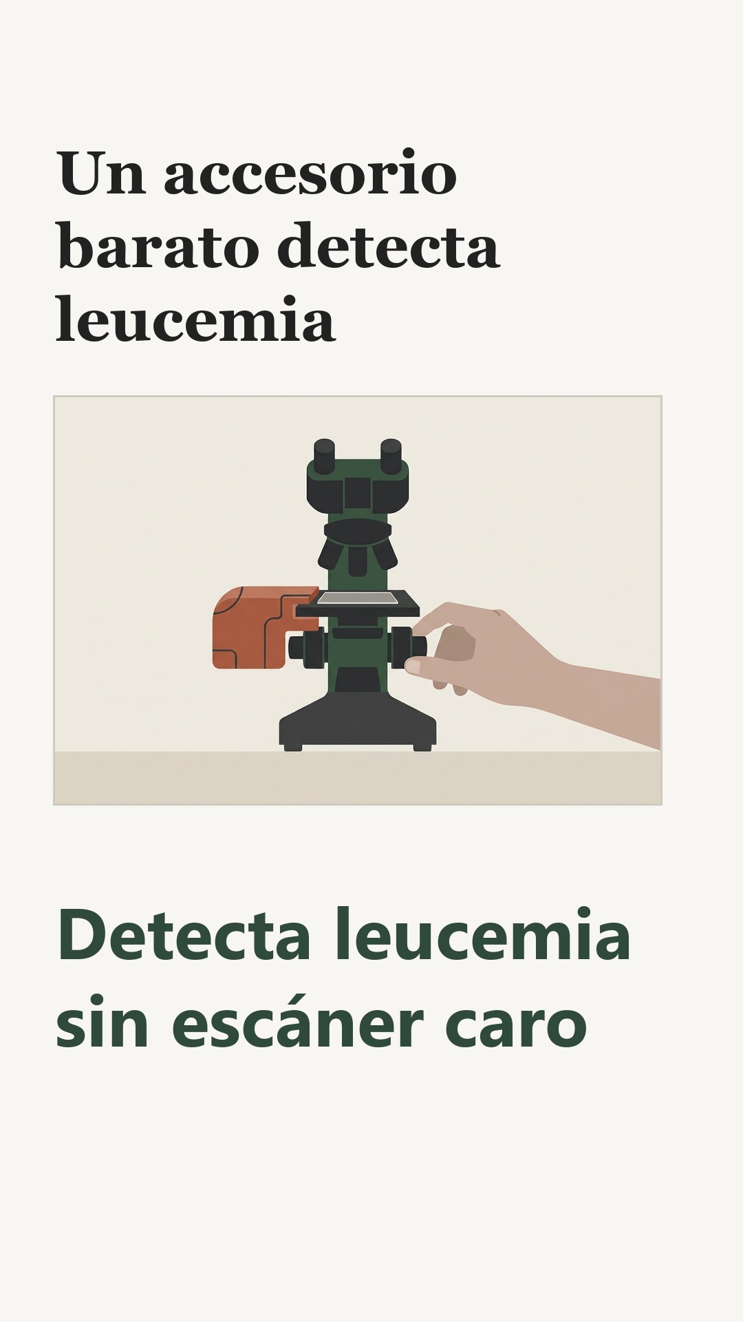
Una caja de piezas impresas en 3D y dos motores pequeños, atornillada a las perillas de un microscopio corriente. El aparato mueve la lámina por su cuenta y una cámara va tomando fotos de la médula ósea extendida sobre el vidrio. El sistema se llama ALLocate y cuesta 150 dólares, sin contar el microscopio.
Un grupo de investigadores de varios hospitales de Estados Unidos lo entrenó con más de 11,000 regiones de médula y 130,000 células. Todo ese material salió de San Francisco. Luego lo probaron con láminas de un segundo hospital que el sistema nunca había visto, en Nueva York.
En 165 láminas de vidrio reales acertó el 88% de los casos, comparado con el diagnóstico del patólogo. En las láminas del primer hospital acertó el 97%; en las del segundo, el 86%. El sistema tardó menos de un minuto en revisar 186 regiones de un hospital externo. Un hematólogo necesita más de cuatro horas para contar células blásticas en 200 regiones.
Hay que decir lo que el aparato no hace. No enfoca hacia arriba y hacia abajo por sí solo, y sin ayuda de una persona el foco puede variar entre una región y otra. Solo separa la leucemia mieloide aguda de una médula normal, y a veces confunde glóbulos blancos jóvenes con células leucémicas. Por eso está pensado para señalar casos a un especialista, no para dar el diagnóstico.
Tampoco hay pruebas de que mejore la salud de los pacientes. El estudio revisó láminas guardadas, no acompañó a nadie durante su tratamiento. No se hizo en África subsahariana ni en países de ingresos bajos, aunque sus autores lo proponen para esos lugares. Y los 165 vidrios vienen de dos hospitales. Los autores esperan que el sistema funcione en otros lugares, pero dicen que hacen falta estudios más grandes en varios centros para confirmarlo.
El ahorro sí es concreto. El artículo describe un estudio en el que los escáneres que suelen necesitar estos programas cuestan unos 300,000 dólares.
Vale la pena preguntar en el hospital público o en la escuela de medicina más cercana si han probado algún accesorio barato de este tipo para revisar sangre o médula. Y conviene preguntar quién revisa el resultado antes de que se actúe sobre él.
Qué significa para usted
El ahorro es real y está medido: el aparato cuesta 150 dólares y los escáneres que suelen usarse cuestan unos 300,000. Pero solo se probó con láminas guardadas de dos hospitales de Estados Unidos, así que todavía no puede decirse que ayude a nadie a vivir más. Pregunte en su hospital o en la escuela de medicina cercana si alguien revisa a mano el resultado antes de que se actúe sobre él.
Yan, E. S., Sun, S., Yin, Z. et al. (2026). An AI-powered self-driving microscope for low-cost acute leukemia detection. Nature Communications. https://doi.org/10.1038/s41467-026-77130-x
Quién pagó: El trabajo recibió apoyo parcial del National Institutes of Health/National Cancer Institute mediante la subvención de apoyo al centro oncológico P30 CA008748, con apoyo adicional del Memorial Sloan Kettering Cancer Center y la Warren Alpert Foundation; el artículo no indica si los financiadores tuvieron alguna influencia, y señala que uno de los autores es accionista de HemeAI, Inc. y otro es empleado de Intuitive Surgical, Inc., empresa que, según el artículo, no tuvo ningún papel en el estudio.
No tome esto como consejo médico profesional.
Los hallazgos de otros estudios que aquí se mencionan provienen de este documento; esos estudios no se consultaron uno por uno.
Versión detalladaLos pasajes copiados del artículo, las ilustraciones y cada fuente con cuánto leímos de ella · 1414 palabras · unos 7 minLeerla →Cerrar
Un aditamento impreso en 3D mueve solo el microscopio y la IA lee las células
Un aditamento barato, probado en 165 láminas de dos hospitales de Estados Unidos, distingue médula ósea con leucemia de médula normal. Todavía no reemplaza a nadie.
En un laboratorio de hematología, alguien se sienta frente al microscopio y cuenta células a ojo: doscientas, trescientas, hasta quinientas por lámina. Busca los blastos, las células inmaduras que delatan una leucemia mieloide aguda. Es lento, cansa y dos personas pueden contar distinto la misma lámina. Un equipo de investigadores de Estados Unidos presentó un sistema que hace buena parte de ese trabajo sin escáner de láminas completas: ALLocate, un aditamento que se monta sobre un microscopio común y que costó unos 150 dólares, o 1,150 con todo y microscopio1.
Piense en lo que cuesta mirar una lámina. El artículo describe un estudio en el que los escáneres que digitalizan un portaobjetos entero, los que usan casi todos los sistemas de inteligencia artificial para leucemia, superan con frecuencia los 300,000 dólares y no están al alcance de muchos hospitales ni de clínicas pequeñas2. Ahí está el problema de fondo: la herramienta que alimenta a la IA es la misma que impide usarla donde más falta hace.
La necesidad no es poca. El artículo describe un estudio en el que la leucemia mieloide aguda representa alrededor del 28% de las muertes por leucemia en el mundo, y el diagnóstico exige identificar al menos 20% de blastos en médula ósea o en sangre, revisando de 200 a 500 células por extendido. El artículo describe un estudio en el que hay países de África subsahariana con 0.1 a 1.3 hematopatólogos por millón de habitantes3. Un especialista por cada millón de personas, en el mejor de los casos: ahí una lámina puede esperar semanas.
El sistema tiene tres piezas. La primera es mecánica: un escáner de microscopio programable y barato, hecho con piezas impresas en 3D, que recorre la lámina solo y toma las fotos. La segunda es un programa que clasifica cada zona de la lámina como útil, sangre o coágulo, para no perder tiempo contando células donde no hay qué contar. La tercera es un detector de células entrenado para distinguir blastos de células normales4.
Los resultados están en el artículo publicado en Nature Communications el 27 de agosto de 2026. Para identificar zonas útiles, el sistema superó 0.99 en una medida que va de 0 a 1 y donde más alto es mejor. Para detectar células, obtuvo 0.90 en la medida estándar de la detección por objetos. En las 165 láminas de vidrio que pasaron por el aparato completo, acertó el 88% de los diagnósticos a nivel de lámina, sin escáner5. En el grupo interno de San Francisco acertó 17 de 18 casos de leucemia y 18 de 18 normales, 97% en total6. En el grupo externo de Nueva York acertó 53 de 56 casos de leucemia y 58 de 73 normales, 86%7.
Hay un dato que cualquier persona que haya esperado un resultado entiende de golpe. En 200 regiones, el sistema tardó en promedio menos de un minuto; un hematopatólogo tardó más de cuatro horas en contar lo mismo a mano8. La diferencia no es de comodidad: es la diferencia entre un resultado el mismo día y un resultado la semana siguiente.
Lo curioso es que este tipo de aparato ya se había probado antes, pero en otra cosa. El artículo describe un estudio en el que un sistema autónomo de microscopía de 250 a 500 dólares detectaba parásitos en sangre periférica, entre ellos los del paludismo9. Y el artículo describe un estudio en el que, en médula ósea, redes neuronales entrenadas con más de 170,000 imágenes de células individuales habían logrado buen desempeño clasificando tipos celulares10. Lo que faltaba era juntar las dos cosas: el aparato barato que mueve la lámina y la IA que decide qué hay en ella.
Conviene ver el tamaño real de la prueba. Fueron 165 láminas de vidrio: 36 de San Francisco y 129 de Nueva York, cada una de un paciente distinto, más grupos de láminas digitalizadas y 186 regiones externas. Los modelos se entrenaron solo con datos de San Francisco y se probaron en Nueva York, lo cual es una prueba más dura que probar en casa. Aun así, 165 láminas no son un servicio clínico.
El aparato tiene límites que importan. No enfoca solo: mueve la lámina de lado a lado, pero el enfoque de arriba abajo lo tiene que hacer una persona, y sin eso la imagen puede salir borrosa en algunas zonas11. A veces confunde granulocitos inmaduros con blastos, sobre todo en médulas reactivas, cuando esas células muestran nucléolos prominentes, un rasgo que se parece al de los blastos leucémicos12. Y no es un diagnóstico: el artículo lo describe como un sistema de apoyo para tamizaje, cuyos resultados revisa un clínico13. No sustituye al patólogo.
Los autores lo dicen con claridad: falta el enfoque automático y falta una interfaz para ver los casos y revisarlos, y el número de láminas físicas todavía es limitado14. También hay que decir quién pagó y quién tiene intereses. El estudio recibió apoyo en parte del Instituto Nacional del Cáncer de Estados Unidos; uno de los autores supervisores tiene acciones en una empresa de IA hematológica y otro trabaja en Intuitive Surgical, que no tuvo papel en el estudio. Los datos completos de entrenamiento no son públicos por privacidad de los pacientes.
Así lo lee weeklyAI: la escena que hay que imaginar no es la del aparato que decide, sino la de la clínica con un solo técnico y una lámpara de escritorio. Ahí, un aditamento de 150 dólares puede volverse la única mirada sobre la lámina. El riesgo no está en que la máquina se equivoque: está en que nadie vuelva a contar células a ojo para comprobarla. Esta lectura se equivocaría si el personal sigue revisando una muestra de láminas al azar y manda siempre los casos dudosos con un especialista, aunque el aparato ya haya dado su número.
También hay que mirar quién responde. Un hospital con pocos especialistas podría comprar el aditamento y presentarlo como si resolviera el diagnóstico completo, sin aclarar que solo señala regiones y cuenta células. La pregunta que cualquier persona puede hacer es concreta: ¿quién firma el resultado y qué pasa si el aparato se equivoca? Si le dicen que el sistema "ya decide solo", vale la pena pedir que un profesional confirme. Y si un equipo así llega a su comunidad, pregunte quién lo va a mantener, quién enseña a usarlo y cómo se comprobará que sigue funcionando meses después.
Aquí weeklyAI se detiene en un detalle: el estudio no se hizo en América Latina, ni en África subsahariana, ni en clínicas de bajos recursos. Se hizo en dos centros oncológicos de Estados Unidos con láminas bien preparadas. Eso no le quita mérito a lo que midió; solo dice que llevarlo a otro lado es otra historia, con otras láminas, otros microscopios y otro personal.
Para el lector de México o de cualquier país de la región, la pregunta útil no es si el aparato es bueno, sino si existe algo parecido en su hospital. La leucemia mieloide aguda se diagnostica con una punción de médula ósea y la revisión de un hematopatólogo; donde no lo hay, el estudio sugiere que un aditamento barato podría ayudar a separar lo urgente de lo que puede esperar. Eso es tamizaje, no diagnóstico. Nadie debe retrasar una consulta por esperar a que llegue la tecnología.
La familia de historias a la que pertenece esta es la de la IA que abarata lo que antes solo estaba en los grandes hospitales. Es la misma lógica del electrocardiógrafo con IA que detecta corazones débiles en la consulta del primer nivel: la herramienta no reemplaza al cardiólogo, pero hace que llegue antes quien lo necesita. La diferencia es que aquí todavía no hay una versión aprobada para usarse en clínicas, ni estudios en pacientes fuera de dos hospitales de Estados Unidos.
Lo que el lector puede llevarse es una pregunta para la próxima vez que oiga de un aparato que "detecta leucemia solo": ¿en cuántas láminas se probó, de cuántos hospitales, y quién revisa lo que dice? Con eso en la mano, la lámpara de escritorio junto al microscopio deja de ser una promesa y se vuelve una herramienta que se puede exigir bien usada.
¿Quién firma el resultado que le entrega un aparato?
De dónde sale cada dato de contexto, y cuánto leímos de cada documento
- Yan, E. S., Sun, S., Yin, Z. et al. (2026). An AI-powered self-driving microscope for low-cost acute leukemia detection. Nature Communications. https://doi.org/10.1038/s41467-026-77130-x - el artículo del que trata esta nota — el artículo completo — el pasaje: "The ALLocate cost ($150, $1150 with microscope) is significantly lower than WSI scanners."
- Yan, E. S., Sun, S., Yin, Z. et al. (2026). An AI-powered self-driving microscope for low-cost acute leukemia detection. Nature Communications. https://doi.org/10.1038/s41467-026-77130-x - el artículo del que trata esta nota — el artículo completo — el pasaje: "While these studies have shown encouraging results, most rely on expensive high-end whole-slide imaging (WSI) systems, which are prohibitively expensive, often exceeding $300,000 USD, and are not readily available in many hospitals or small clinics"
- Yan, E. S., Sun, S., Yin, Z. et al. (2026). An AI-powered self-driving microscope for low-cost acute leukemia detection. Nature Communications. https://doi.org/10.1038/s41467-026-77130-x - el artículo del que trata esta nota — el artículo completo — el pasaje: "As clinical volumes rise, the need for faster, standardized diagnostics grows, especially in resource-constrained settings with limited access to expert hematologists or pathologists. For instance, countries in sub-Saharan Africa have only 0.1 to 1.3 hematopathologists per million people, creating substantial barriers to the timely diagnosis of hematologic malignancies."
- Yan, E. S., Sun, S., Yin, Z. et al. (2026). An AI-powered self-driving microscope for low-cost acute leukemia detection. Nature Communications. https://doi.org/10.1038/s41467-026-77130-x - el artículo del que trata esta nota — el artículo completo — el pasaje: "It integrates an inexpensive, 3D-printed programmable microscope scanner with two deep learning modules: a CNN-based region classifier and a YOLOv8-based, one-step cell detection and classification model."
- Yan, E. S., Sun, S., Yin, Z. et al. (2026). An AI-powered self-driving microscope for low-cost acute leukemia detection. Nature Communications. https://doi.org/10.1038/s41467-026-77130-x - el artículo del que trata esta nota — el artículo completo — el pasaje: "ALLocate achieves an area under the receiver operating characteristic curve greater than 0.99 for region-of-interest identification, a mean average precision at 50% intersection over union of 0.90 for cell detection, and 88% accuracy for slide-level diagnosis on glass slides without requiring a whole-slide scanner."
- Yan, E. S., Sun, S., Yin, Z. et al. (2026). An AI-powered self-driving microscope for low-cost acute leukemia detection. Nature Communications. https://doi.org/10.1038/s41467-026-77130-x - el artículo del que trata esta nota — el artículo completo — el pasaje: "On the internal UCSF cohort, ALLocate correctly classified 17 out of 18 AML cases (94.4%) and 18 out of 18 normal cases (100%), corresponding to an overall accuracy of 97%."
- Yan, E. S., Sun, S., Yin, Z. et al. (2026). An AI-powered self-driving microscope for low-cost acute leukemia detection. Nature Communications. https://doi.org/10.1038/s41467-026-77130-x - el artículo del que trata esta nota — el artículo completo — el pasaje: "Importantly, performance remained robust on the fully independent external MSKCC cohort, where ALLocate correctly classified 53 out of 56 AML cases (94.6%) and 58 out of 73 normal cases (79.5%), achieving an overall accuracy of 86.0%."
- Yan, E. S., Sun, S., Yin, Z. et al. (2026). An AI-powered self-driving microscope for low-cost acute leukemia detection. Nature Communications. https://doi.org/10.1038/s41467-026-77130-x - el artículo del que trata esta nota — el artículo completo — el pasaje: "However, the evaluation time vastly differed, with ALLocate requiring, on average, less than 1 minute for evaluation compared to more than 4 hours for a hematopathologist to manually perform blast counting in 200 regions."
- Yan, E. S., Sun, S., Yin, Z. et al. (2026). An AI-powered self-driving microscope for low-cost acute leukemia detection. Nature Communications. https://doi.org/10.1038/s41467-026-77130-x - el artículo del que trata esta nota — el artículo completo — el pasaje: "For example, Li et al. developed an autonomous, low-cost microscopy system (~$250–$500) capable of detecting infectious agents in peripheral blood smears, including Loa loa , Trypanosoma brucei rhodesiense , and malaria"
- Yan, E. S., Sun, S., Yin, Z. et al. (2026). An AI-powered self-driving microscope for low-cost acute leukemia detection. Nature Communications. https://doi.org/10.1038/s41467-026-77130-x - el artículo del que trata esta nota — el artículo completo — el pasaje: "Matek et al. applied convolutional neural networks (CNNs) to over 170,000 single-cell images from marrow smears, achieving high performance across cell types"
- Yan, E. S., Sun, S., Yin, Z. et al. (2026). An AI-powered self-driving microscope for low-cost acute leukemia detection. Nature Communications. https://doi.org/10.1038/s41467-026-77130-x - el artículo del que trata esta nota — el artículo completo — el pasaje: "First, while the current hardware enables automated XY stage scanning for slide pre-screening, it lacks automated Z-axis focusing, which, without human input, may result in focal variability across regions."
- Yan, E. S., Sun, S., Yin, Z. et al. (2026). An AI-powered self-driving microscope for low-cost acute leukemia detection. Nature Communications. https://doi.org/10.1038/s41467-026-77130-x - el artículo del que trata esta nota — el artículo completo — el pasaje: "Finally, we observed occasional misclassification of immature granulocytes as blasts, particularly in reactive marrow conditions. These errors most frequently occurred when immature granulocytes exhibited prominent nucleoli, a feature that can overlap morphologically with leukemic blasts and challenge the model’s discriminative capacity."
- Yan, E. S., Sun, S., Yin, Z. et al. (2026). An AI-powered self-driving microscope for low-cost acute leukemia detection. Nature Communications. https://doi.org/10.1038/s41467-026-77130-x - el artículo del que trata esta nota — el artículo completo — el pasaje: "In practical settings, ALLocate operates as an assistive system rather than a standalone diagnostic replacement: detected regions and cell counts are reviewed in aggregate, and outputs can be verified by a clinician."
- Yan, E. S., Sun, S., Yin, Z. et al. (2026). An AI-powered self-driving microscope for low-cost acute leukemia detection. Nature Communications. https://doi.org/10.1038/s41467-026-77130-x - el artículo del que trata esta nota — el artículo completo — el pasaje: "Future work will focus on incorporating autofocus functionality and developing a unified interface to facilitate clinical integration. Also, although glass-level validation was performed on both the UCSF dataset (including an independent test set) and the MSKCC external validation cohort, the overall number of physical glass slides remains relatively limited."
Yan, E. S., Sun, S., Yin, Z. et al. (2026). An AI-powered self-driving microscope for low-cost acute leukemia detection. Nature Communications. https://doi.org/10.1038/s41467-026-77130-x
Quién pagó: El trabajo recibió apoyo parcial del National Institutes of Health/National Cancer Institute mediante la subvención de apoyo al centro oncológico P30 CA008748, con apoyo adicional del Memorial Sloan Kettering Cancer Center y la Warren Alpert Foundation; el artículo no indica si los financiadores tuvieron alguna influencia, y señala que uno de los autores es accionista de HemeAI, Inc. y otro es empleado de Intuitive Surgical, Inc., empresa que, según el artículo, no tuvo ningún papel en el estudio.
No tome esto como consejo médico profesional.
Los hallazgos de otros estudios que aquí se mencionan provienen de este documento; esos estudios no se consultaron uno por uno.
experiment · Nature communications · the paper, 27 Aug 2026 · free
A $150 Box That Moves the Microscope, and Finds Leukemia Cells
On 165 glass slides from two US hospitals, an add-on called ALLocate named the right answer 88 times in 100. It cannot focus itself, and a specialist still checks its work.
Short version · the longer version follows, about 5 min
Ask the weeklyAI chat
Ask me about AI in your life: I point you to the stories weeklyAI has published on it, this week and before.
Conversations are saved for as long as weeklyAI exists, to improve the publication. Answers come in the language you write in.
- The study at a glance
- Who
- Bone marrow smear slides from patients at two US cancer centers
- How many
- 165 physical glass slides
- Where
- United States (San Francisco and New York)
- When
- 2026
- Kind of study
- experiment
- Who did it
- University of California, San Francisco and Memorial Sloan Kettering Cancer Center
- The limit that matters
- It cannot focus itself, and a specialist must still review its findings.
These are shares of cases called correctly by the system on physical glass slides; the second hospital's slides were never seen during training, and the authors say the number of glass slides tested is still small.
Time to count blasts in 200 regions
Under a minute for the system, more than four hours for the specialist
It is a screening aid, not a diagnosis, and it has so far been tested on slides from two US cancer centers, with the authors calling for larger studies across more centers.
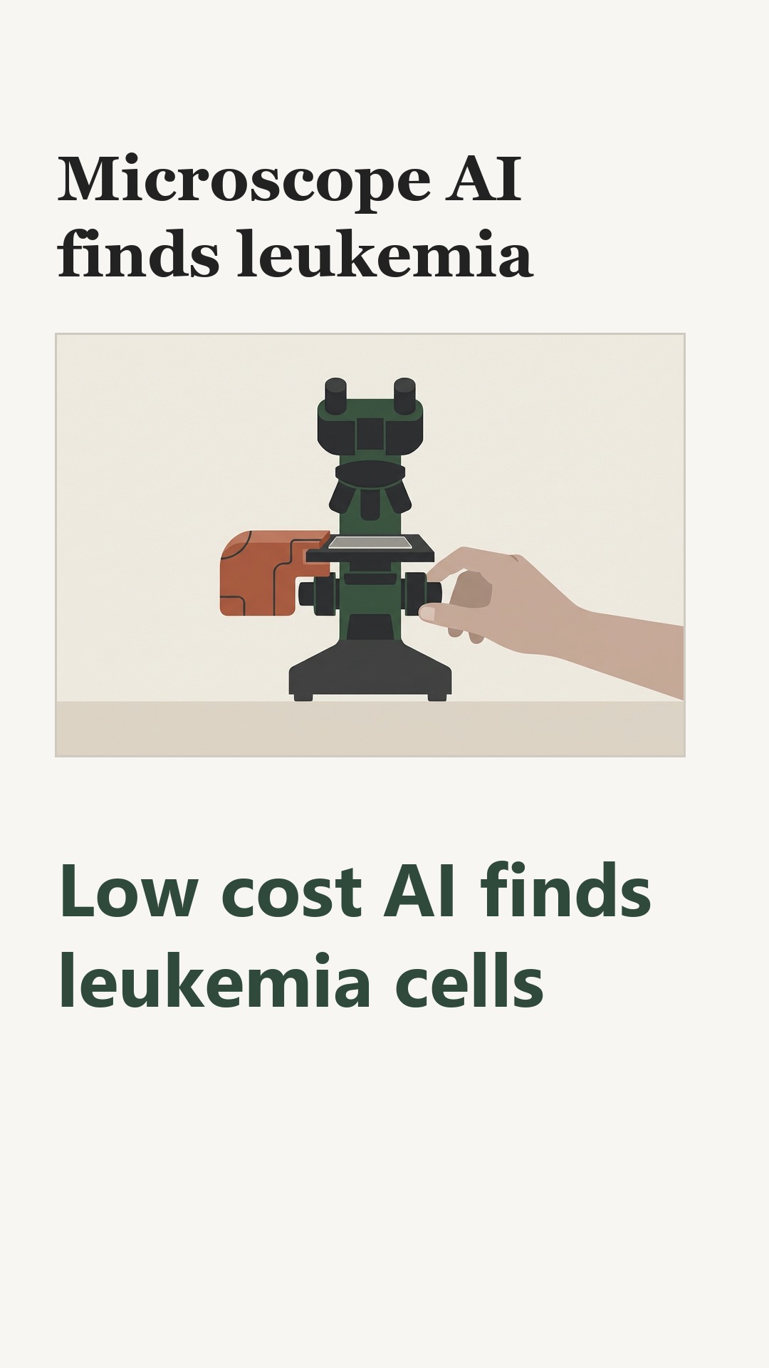
Two small motors, a 3D-printed shell and an Arduino board, bolted onto the knobs that move an ordinary microscope's stage. The slide travels on its own, and a camera takes pictures of each patch of bone marrow.
Downstream, two programs sort the pictures. One picks the patches worth reading. The other circles each cell and marks it as a blast or a normal cell. A high share of blasts points to acute myeloid leukemia. The article says this disease accounts for about 28 in 100 leukemia deaths worldwide.
Researchers at the University of California, San Francisco, trained the programs on their own slides. One program learned from about 9,000 marrow regions. The other learned from nearly 110,000 cells. They tested the programs on about 2,300 more regions and about 27,000 more cells. Then they tested the system at Memorial Sloan Kettering Cancer Center in New York, on slides it had never seen.
On 165 real glass slides, the system called 88 of every 100 cases correctly. At UCSF it got 97 in 100; at the second hospital, 86. On 200 regions from the second hospital, counting blasts took it under a minute. A hematologist needed more than four hours to count the same regions by hand.
The device does not focus up and down by itself, so a person must still handle the microscope. It separates AML from normal marrow and nothing else. It sometimes mistakes immature granulocytes for blasts, mostly in marrow that is reacting to other illness. The article describes it as a screening aid whose findings an expert reviews. Whether patients actually fare better was not measured.
The add-on costs about $150, or $1,150 with a microscope, against roughly $300,000 for the whole-slide scanners such software usually needs.
Look for whether a public or teaching hospital near you has tried a low-cost microscope add-on for blood or marrow screening, and ask who reviews its findings before any result reaches a patient.
What this means for you
The study took place at two cancer centers in the United States, on marrow slides read after the fact, so nothing here yet describes a clinic near you. What you can ask is who reviews any automated result before it reaches a patient. Watch for a hospital testing a cheap microscope add-on; the claim to weigh is that a specialist still checks its work.
Yan, E. S., Sun, S., Yin, Z. et al. (2026). An AI-powered self-driving microscope for low-cost acute leukemia detection. Nature Communications. https://doi.org/10.1038/s41467-026-77130-x
Who paid: The work was supported in part by the National Institutes of Health/National Cancer Institute Cancer Center Support Grant P30 CA008748, with additional support from Memorial Sloan Kettering Cancer Center and the Warren Alpert Foundation; the article does not say whether funders had any say, and notes that one author is an equity holder of HemeAI, Inc. and another is an employee of Intuitive Surgical, Inc., which the article says had no role in the study.
Do not take this as professional medical advice.
The findings of other studies mentioned here come from this document; those studies were not opened one by one.
The longer versionThe passages copied from the paper, the pictures, and every source with how much of it we read · 917 words · about 5 minRead it →Close
A $150 Add-On Turns an Ordinary Microscope Into a Leukemia Screener
In bone marrow slides from two US cancer centers, the system flagged acute myeloid leukemia with 88% accuracy on real glass slides. It is a screening aid, not a diagnosis, and it has so far been tested on slides from two US cancer centers, with the authors calling for larger studies across more centers.
Consider what a pathologist does with a bone marrow smear. Under a light microscope, the smear is scanned at low power for the patches where the white-cell precursors cluster, and then, in those patches, several hundred cells are counted by eye. A diagnosis of acute myeloid leukemia, or AML, rests on a share of those cells: at least one in five must be a leukemic blast.
ALLocate, described in Nature Communications by Ethan S. Yan, Shenghuan Sun and colleagues, splits that job in two. A 3D-printed rig of two small motors clamps onto the stage knobs of an ordinary microscope and moves the slide by itself. A first program sorts the captured images into clotted, blood-filled and usable regions; a second, built on the YOLOv8 object detector, finds and labels individual cells as blasts or normal cells. The rig costs $150, or $1,150 with the microscope included1.
The authors trained their models only on slides from the University of California, San Francisco, then tested them on slides from UCSF and from Memorial Sloan Kettering Cancer Center. On 165 real glass slides run through the complete system, the system's overall accuracy was 88%: 97% on the 36 UCSF slides and 86% on the 129 from Sloan Kettering234. The Sloan Kettering result rests on 53 of 56 AML slides called correctly and 58 of 73 normal slides4. No margin of error was reported for the overall figure.
On the 40 Sloan Kettering slides that were scanned digitally, the system told every AML slide apart from every normal one under an ideal cutoff. Even when held to the textbook cutoffs of 5% blasts for benign and 20% for AML, the system achieved 93% accuracy. On the cell-counting task it reached a mean average precision of 0.90, and on finding the right regions to look at, a score above 0.992. Counting 200 regions took it under a minute, against more than four hours for a hematopathologist5.
The limits are the authors' own. The rig moves the slide side to side but cannot focus up and down on its own, so a person must still handle that part6. It sometimes mistakes immature granulocytes, a normal marrow cell, for blasts, especially in reactive marrow7. It was built to tell AML from normal marrow only, and the authors describe it as an assistive system whose output a clinician reviews, not a replacement for diagnosis8. The number of glass slides tested is small, and no case-management screen for pathologists exists yet9.
The article describes a study in which the scanners that most AI leukemia tools depend on cost more than $300,000 and are scarce in small hospitals and clinics10. The article describes a study in which sub-Saharan Africa has between 0.1 and 1.3 hematopathologists per million people11. A tool that attaches to a microscope already in the building is a different proposition from a machine that has to be bought.
Others have tried pieces of this. The article describes a study in which a low-cost autonomous microscope in the $250–$500 range detected malaria and two parasitic worms in blood smears12. The article describes a study in which convolutional networks trained on more than 170,000 single marrow cells classified cell types with high performance13. ALLocate's claim is narrower and more practical: one stage of detection, on a microscope someone already owns.
Here is weeklyAI's reading. A new tool rarely takes the expert out of the room; it changes who waits and for how long. Expect a clinic that adopts this to still send every flagged slide to a hematopathologist for sign-off, so the visible gain is a shorter wait for that expert's attention. This reading would be wrong if clinics with no such expert began treating the blast percentage as a stand-alone result.
The price comparison will be the loudest thing in most retellings, and it should not be. A scanner costing a fortune beside a rig costing $150 makes the problem look solved. Ask instead what the 88% means for the roughly one in eight slides the system got wrong, and in which direction those errors fell: a sick person sent home, or a healthy person sent for more tests.
And ask the plain question about the building. A working microscope and someone trained to use it are the floor this system stands on. Where both exist, a cheap add-on has somewhere to land; where neither does, it does not.
For a family in the United States, Canada or Latin America, the honest position is this: nothing here changes care today, and no one should skip or delay a test on the strength of it. What a reader can ask for is smaller and more useful. At a public clinic or a hospital lab, ask whether the pathology service has a microscope and a trained operator, and whether AI tools are being reviewed by a named specialist before any result is acted on. The question costs nothing, and it tells you more than any accuracy figure in a headline.
The slide under the lens still needs a person at the eyepiece. What may change is how long it sits there.
Where each piece of context comes from, and how much of it we read
- Yan, E. S., Sun, S., Yin, Z. et al. (2026). An AI-powered self-driving microscope for low-cost acute leukemia detection. Nature Communications. https://doi.org/10.1038/s41467-026-77130-x - the article this story is about — the whole article — the passage: "The ALLocate cost ($150, $1150 with microscope) is significantly lower than WSI scanners."
- Yan, E. S., Sun, S., Yin, Z. et al. (2026). An AI-powered self-driving microscope for low-cost acute leukemia detection. Nature Communications. https://doi.org/10.1038/s41467-026-77130-x - the article this story is about — the whole article — the passage: "ALLocate achieves an area under the receiver operating characteristic curve greater than 0.99 for region-of-interest identification, a mean average precision at 50% intersection over union of 0.90 for cell detection, and 88% accuracy for slide-level diagnosis on glass slides without requiring a whole-slide scanner."
- Yan, E. S., Sun, S., Yin, Z. et al. (2026). An AI-powered self-driving microscope for low-cost acute leukemia detection. Nature Communications. https://doi.org/10.1038/s41467-026-77130-x - the article this story is about — the whole article — the passage: "On the internal UCSF cohort, ALLocate correctly classified 17 out of 18 AML cases (94.4%) and 18 out of 18 normal cases (100%), corresponding to an overall accuracy of 97%."
- Yan, E. S., Sun, S., Yin, Z. et al. (2026). An AI-powered self-driving microscope for low-cost acute leukemia detection. Nature Communications. https://doi.org/10.1038/s41467-026-77130-x - the article this story is about — the whole article — the passage: "Importantly, performance remained robust on the fully independent external MSKCC cohort, where ALLocate correctly classified 53 out of 56 AML cases (94.6%) and 58 out of 73 normal cases (79.5%), achieving an overall accuracy of 86.0%."
- Yan, E. S., Sun, S., Yin, Z. et al. (2026). An AI-powered self-driving microscope for low-cost acute leukemia detection. Nature Communications. https://doi.org/10.1038/s41467-026-77130-x - the article this story is about — the whole article — the passage: "However, the evaluation time vastly differed, with ALLocate requiring, on average, less than 1 minute for evaluation compared to more than 4 hours for a hematopathologist to manually perform blast counting in 200 regions."
- Yan, E. S., Sun, S., Yin, Z. et al. (2026). An AI-powered self-driving microscope for low-cost acute leukemia detection. Nature Communications. https://doi.org/10.1038/s41467-026-77130-x - the article this story is about — the whole article — the passage: "First, while the current hardware enables automated XY stage scanning for slide pre-screening, it lacks automated Z-axis focusing, which, without human input, may result in focal variability across regions."
- Yan, E. S., Sun, S., Yin, Z. et al. (2026). An AI-powered self-driving microscope for low-cost acute leukemia detection. Nature Communications. https://doi.org/10.1038/s41467-026-77130-x - the article this story is about — the whole article — the passage: "Finally, we observed occasional misclassification of immature granulocytes as blasts, particularly in reactive marrow conditions. These errors most frequently occurred when immature granulocytes exhibited prominent nucleoli, a feature that can overlap morphologically with leukemic blasts and challenge the model’s discriminative capacity."
- Yan, E. S., Sun, S., Yin, Z. et al. (2026). An AI-powered self-driving microscope for low-cost acute leukemia detection. Nature Communications. https://doi.org/10.1038/s41467-026-77130-x - the article this story is about — the whole article — the passage: "In practical settings, ALLocate operates as an assistive system rather than a standalone diagnostic replacement: detected regions and cell counts are reviewed in aggregate, and outputs can be verified by a clinician."
- Yan, E. S., Sun, S., Yin, Z. et al. (2026). An AI-powered self-driving microscope for low-cost acute leukemia detection. Nature Communications. https://doi.org/10.1038/s41467-026-77130-x - the article this story is about — the whole article — the passage: "Future work will focus on incorporating autofocus functionality and developing a unified interface to facilitate clinical integration. Also, although glass-level validation was performed on both the UCSF dataset (including an independent test set) and the MSKCC external validation cohort, the overall number of physical glass slides remains relatively limited."
- Yan, E. S., Sun, S., Yin, Z. et al. (2026). An AI-powered self-driving microscope for low-cost acute leukemia detection. Nature Communications. https://doi.org/10.1038/s41467-026-77130-x - the article this story is about — the whole article — the passage: "While these studies have shown encouraging results, most rely on expensive high-end whole-slide imaging (WSI) systems, which are prohibitively expensive, often exceeding $300,000 USD, and are not readily available in many hospitals or small clinics"
- Yan, E. S., Sun, S., Yin, Z. et al. (2026). An AI-powered self-driving microscope for low-cost acute leukemia detection. Nature Communications. https://doi.org/10.1038/s41467-026-77130-x - the article this story is about — the whole article — the passage: "As clinical volumes rise, the need for faster, standardized diagnostics grows, especially in resource-constrained settings with limited access to expert hematologists or pathologists. For instance, countries in sub-Saharan Africa have only 0.1 to 1.3 hematopathologists per million people, creating substantial barriers to the timely diagnosis of hematologic malignancies."
- Yan, E. S., Sun, S., Yin, Z. et al. (2026). An AI-powered self-driving microscope for low-cost acute leukemia detection. Nature Communications. https://doi.org/10.1038/s41467-026-77130-x - the article this story is about — the whole article — the passage: "For example, Li et al. developed an autonomous, low-cost microscopy system (~$250–$500) capable of detecting infectious agents in peripheral blood smears, including Loa loa , Trypanosoma brucei rhodesiense , and malaria"
- Yan, E. S., Sun, S., Yin, Z. et al. (2026). An AI-powered self-driving microscope for low-cost acute leukemia detection. Nature Communications. https://doi.org/10.1038/s41467-026-77130-x - the article this story is about — the whole article — the passage: "Matek et al. applied convolutional neural networks (CNNs) to over 170,000 single-cell images from marrow smears, achieving high performance across cell types"
Yan, E. S., Sun, S., Yin, Z. et al. (2026). An AI-powered self-driving microscope for low-cost acute leukemia detection. Nature Communications. https://doi.org/10.1038/s41467-026-77130-x
Who paid: The work was supported in part by the National Institutes of Health/National Cancer Institute Cancer Center Support Grant P30 CA008748, with additional support from Memorial Sloan Kettering Cancer Center and the Warren Alpert Foundation; the article does not say whether funders had any say, and notes that one author is an equity holder of HemeAI, Inc. and another is an employee of Intuitive Surgical, Inc., which the article says had no role in the study.
Do not take this as professional medical advice.
The findings of other studies mentioned here come from this document; those studies were not opened one by one.