review of studies · The Indian journal of radiology & imaging · la publicación, 13 ago 2026 · gratis
Un programa de cómputo ayudó a hallar más tumores en Suecia
El sistema redujo la carga de los médicos, pero todavía no demuestra si salva vidas ni cómo funcionaría en otras poblaciones.
Versión breve · la versión detallada sigue, unos 6 min
Pregunte al chat de weeklyAI
Pregúnteme sobre la IA en su vida: le señalo las notas que weeklyAI ha publicado sobre el tema, esta semana y antes.
Las conversaciones se guardan mientras exista weeklyAI, para mejorar la publicación. Se responde en el idioma en que usted escribe.
- El estudio, de un vistazo
- Quiénes
- Mujeres del programa nacional de detección de cáncer de mama
- Cuántos
- 105,934 mujeres
- Dónde
- Suecia
- Cuándo
- 2023 a 2026
- Tipo de estudio
- experiment
- Quién lo hizo
- Departamento de Imagen Mamaria, HealthBay Clinics, Dubái, y programa de detección de Escocia
- El límite que importa
- No hay datos de mortalidad y casi todas las pacientes eran caucásicas de países ricos
Son aumentos de detección de cáncer en distintos estudios y esquemas, no el mismo grupo de mujeres; el primero y el segundo son del mismo ensayo sueco en momentos distintos.
Revisión doble habitual frente a revisión apoyada por el programa de cómputo (ensayo sueco)
6.1 frente a 5.1 por cada 1,000 en el primer análisis; 6.4 frente a 5.0 en el de 2025
Bajaron 12%
Bajó 44.3%
Igual en ambos grupos: 98.5%
Detectar más tumores no significa de inmediato que mueran menos mujeres.
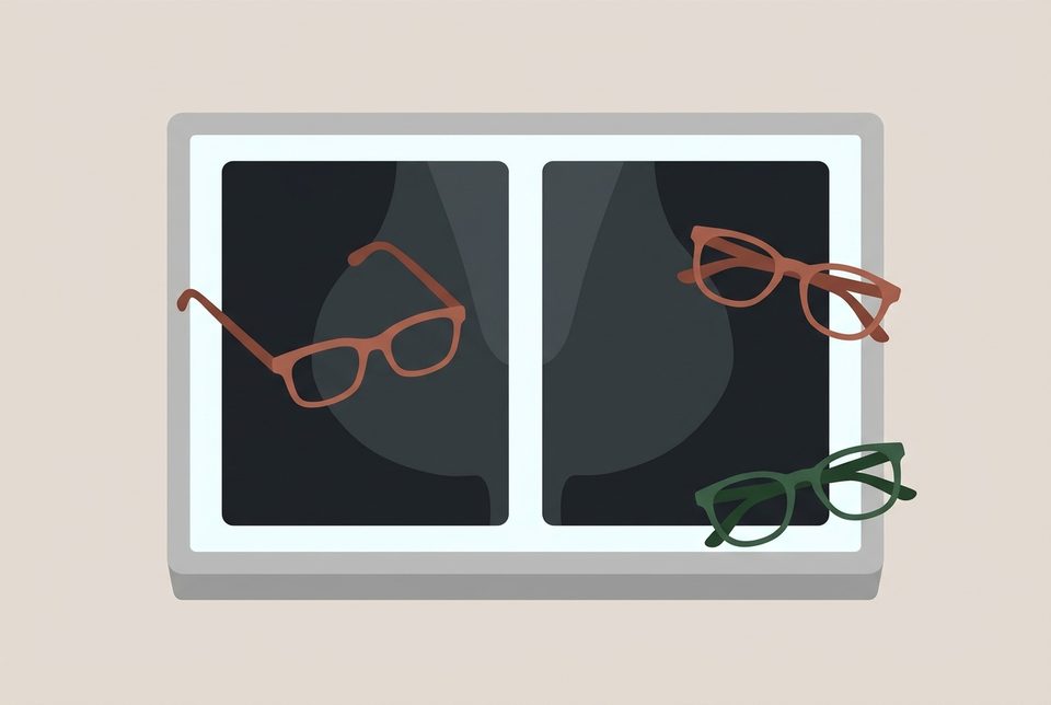
Una sala de lectura en Suecia, miles de mamografías digitales y un sistema de inteligencia artificial que califica cada placa con un puntaje de riesgo. La revisión recoge un estudio en el que participaron 105,934 mujeres de un programa nacional de detección. En ese estudio, la mitad pasó por la revisión habitual con dos radiólogos. La otra mitad entró a un esquema nuevo: si el programa marcaba un riesgo bajo, la imagen la revisaba un solo especialista; si marcaba un riesgo alto, la examinaban dos.
La revisión recoge que ese filtro redujo en 44.3% el trabajo de lectura de las mamografías. Los casos de riesgo bajo los revisó un solo especialista en lugar de dos.
La revisión recoge que el grupo con apoyo del programa detectó 29% más cánceres. La revisión recoge que la mayoría fueron tumores invasivos pequeños que todavía no llegaban a los ganglios. La revisión recoge además que los tumores que aparecieron entre una revisión y otra cayeron 12%. La revisión recoge que el programa no aumentó las falsas alarmas. La revisión recoge que, en el análisis principal, la certeza de que una mama sana saliera limpia fue la misma en ambos grupos, de 98.5%.
La duda empieza con la palabra cáncer. Detectar más tumores no significa de inmediato que mueran menos mujeres. Los casos extra que encontró el sistema fueron sobre todo tumores pequeños que todavía no llegaban a los ganglios. La revisión recoge que entre ellos hubo también más tumores de los llamados agresivos. Ese perfil también se parece al de tumores que nunca habrían causado daño. En algunos casos son tumores que jamás habrían causado molestias ni la muerte, un problema conocido como sobrediagnóstico que puede llevar a biopsias o tratamientos innecesarios.
La revisión señala que ningún ensayo clínico terminado con esta tecnología ha medido aún si reduce la mortalidad a largo plazo.
La revisión recoge que las mujeres del estudio eran suecas y vivían en un país de ingresos altos con un sistema de salud bien financiado. La revisión advierte que en un hospital público de América Latina o en una clínica que atiende a poblaciones más diversas, los programas de cómputo pueden rendir de otra manera si fueron entrenados con imágenes distintas a las de la gente local.
El software no trabajó solo. En este esquema, las imágenes de riesgo bajo las revisó un solo especialista en lugar de dos, y las de riesgo alto siguieron pasando por la revisión habitual de dos. Si en su clínica le ofrecen una mastografía leída con ayuda de inteligencia artificial, la pregunta que vale la pena llevar consigo es sencilla: ¿este programa se probó antes en mujeres de nuestra comunidad?
Qué significa para usted
Ese resultado viene de mujeres suecas dentro de un programa nacional que ya revisaba cada mamografía con dos especialistas. En su clínica, si le hablan de inteligencia artificial, pregunte si el programa se probó antes con mujeres de su comunidad y si la revisión sigue en manos de un profesional. Nadie ha medido todavía si este sistema reduce las muertes por cáncer de mama.
Kumar, A., Lip, G. (2026). Artificial Intelligence in Breast Imaging and Screening: Current Evidence, Applications, and Future Directions. Indian Journal of Radiology and Imaging. https://doi.org/10.1055/s-0046-1827249
Quién pagó: El artículo no indica ninguna fuente de financiación y declara ausencia de conflictos de interés; no dice quién financió el estudio ni quién prestó equipos o programas.
No tome esto como consejo médico profesional.
Los hallazgos de otros estudios que aquí se mencionan los conocemos por este documento, que fue el que leímos; no abrimos cada uno de esos estudios.
Versión detalladaLos pasajes copiados del artículo, las ilustraciones y cada fuente con cuánto leímos de ella · 1277 palabras · unos 6 minLeerla →Cerrar
La IA encontró más cánceres en un programa sueco de mamografías. Lo que aún no se sabe es si eso salva vidas.
Un ensayo aleatorizado de casi 106,000 mujeres midió la detección, la carga de trabajo y los cánceres que aparecen entre una mamografía y otra. La mortalidad no estaba entre lo que se midió.
La revisión recoge un estudio en el que, dentro del programa nacional de detección de cáncer de mama de Suecia, se asignó al azar a 105,934 mujeres a dos formas de revisar sus mamografías: la revisión doble habitual, con dos radiólogos por estudio, o una revisión apoyada por un programa de inteligencia artificial. La revisión recoge un estudio en el que el programa le ponía un puntaje de riesgo a cada estudio: los de riesgo bajo pasaban a un solo lector y los de riesgo alto seguían con los dos. Esa es la configuración que se probó: la IA sustituyó a uno de los dos lectores solo en los casos de menor riesgo1.
Con ese esquema se detectaron más cánceres. La revisión recoge un primer análisis de seguridad, publicado en 2023, en el que la tasa de detección fue de 6.1 por cada 1,000 mujeres examinadas, frente a 5.1 con la revisión doble: un 20% más, sin aumento de los falsos positivos, y con 44.3% menos carga de lectura2. La revisión recoge un análisis de desempeño de 2025 que confirmó un 29% más de detección (6.4 frente a 5.0 por 1,000), sobre todo de cánceres pequeños, invasivos y sin ganglios afectados, e incluyó 27% más de subtipos agresivos3. La revisión recoge un análisis del objetivo principal, en 2026, que reportó una sensibilidad de 80.5% frente a 73.8%, con la misma especificidad de 98.5% en ambos brazos, y una reducción de 12% en los cánceres de intervalo, los que aparecen entre una mamografía y la siguiente4.
Detectar más no es lo mismo que vivir más. El propio artículo lo dice: un aumento en la detección no se traduce automáticamente en menos muertes por cáncer de mama, y los datos de mortalidad no están disponibles en ningún estudio terminado de IA para tamizaje5. La revisión señala que también hay que mirar qué se detectó de más: junto con los cánceres agresivos aparecieron más cánceres luminal A de bajo grado y lesiones in situ, un perfil que merece escrutinio por el riesgo de sobrediagnóstico, es decir, encontrar tumores que nunca habrían causado daño y que llevan a biopsias y tratamientos innecesarios. Y el esquema probado fue una sustitución parcial, no un reemplazo del radiólogo1.
Piense en la sala de lectura de un programa público de tamizaje. La norma europea pide que cada mamografía la lean dos radiólogos, para no dejar pasar nada; el problema es que faltan radiólogos en todo el mundo y esa doble lectura pesa6. La mamografía, además, arrastra tasas altas de falsos positivos y falsos negativos: estudios que asustan sin razón y cánceres que se escapan7. Lo curioso es que el cáncer de mama sigue siendo el más diagnosticado entre las mujeres y una de las principales causas de muerte por cáncer8.
El software no funciona de una sola manera. Puede leerse solo, sin radiólogo; puede entrar como segundo lector, después del primero; puede clasificar por riesgo para decidir quién va a lectura sencilla y quién a doble; o puede declarar normales los casos de bajo riesgo sin que ningún radiólogo los vea1. Cada configuración cambia quién responde cuando algo falla. La revisión que estamos comentando reúne evidencia de las cuatro, y conviene no confundirlas.
Otros programas ya lo probaron. La revisión recoge un estudio de implementación en el programa nacional de Alemania, con más de 460,000 mujeres, que encontró 17.6% más detección con lectura doble apoyada por IA y sin subir la tasa de llamados a repetir el estudio9. La revisión recoge un estudio en Corea del Sur, donde la lectura es de un solo radiólogo, en el que la detección fue de 5.70 frente a 5.01 por 1,000 sin diferencia significativa en los llamados10. La revisión recoge un esquema parcialmente autónomo, en el que los casos de bajo riesgo se dieron por normales sin que los viera un radiólogo, que redujo la carga de lectura 63.6% y subió la detección 15.2%11. Y la revisión recoge una revisión sistemática de 31 estudios y más de dos millones de exámenes que halló que, como segundo lector, la IA mantuvo o subió un poco la sensibilidad; en triage, redujo el volumen de lectura entre 40 y 90% sin perder detección12.
El resumen de una revisión de diez años de evidencia, del que solo pudimos leer el resumen porque el texto completo está detrás de una suscripción, junta 26 estudios publicados entre 2019 y 202513. Su conclusión es sobria: la IA sirve como segundo lector y como herramienta de triage en programas organizados, no como reemplazo autónomo del radiólogo14. Esa misma revisión advierte que el desempeño promedio es alto pero tan disperso que no se puede trasladar a un programa nuevo sin validarlo localmente15, y que las ganancias más grandes vienen de estudios no aleatorizados, justo el patrón que produciría un sesgo16.
Así lo leemos nosotros. Cuando un programa de salud decide quién está sano y quién es sospechoso, la pregunta que suele quedar sin respuesta es con qué casos se entrenó el sistema y a quién deja fuera. El artículo lo admite: solo entre 0 y 25% de los estudios reportaron raza o etnia, casi todas las pacientes eran caucásicas y no hubo estudios en países de bajos ingresos17. La revisión recoge un estudio en el que un modelo entrenado con datos amplios pero poco diversos rindió mucho peor al aplicarse a una población heterogénea de Los Ángeles, con sensibilidades de 0.4 a 0.518. No es un defecto de este ensayo; es algo que conviene vigilar en cualquier programa que adopte estas herramientas.
Hay un asunto de dinero y de personal que el entusiasmo tapa. El ahorro de trabajo de 31 a 64% en programas de doble lectura19 puede servir para sostener la doble lectura donde falta gente, o puede usarse para recortar plantilla. Depende de quién administre. Si el software llega como producto licenciado, su costo puede frenar a los programas públicos. Usted puede preguntar en su centro de salud cuánto cuesta el sistema, quién lo paga y en qué se usa el tiempo que ahorra.
La revisión pide lo que falta: vigilancia después de la venta, datos inclusivos y análisis de costo-efectividad20. Nada de eso está resuelto. Tampoco hay estudios prospectivos de los modelos que calculan el riesgo futuro de una mujer a partir de una mamografía: la revisión recoge dos revisiones sistemáticas que encontraron que todos los estudios incluidos eran retrospectivos, y que la calibración —que el riesgo anunciado coincida con lo que de verdad ocurre— rara vez se reportaba.
Para una familia en México o en cualquier país de la región, la pregunta práctica no es si la IA es buena. Es si el programa donde se hace la mamografía usa estas herramientas, con qué población se probaron y quién revisa los casos que el sistema marca como normales. Pregunte también si el programa lleva registro de errores por grupo de población. Un programa que no puede contestar eso todavía no sabe si la herramienta le funciona a usted.
Esa mamografía que le toca el próximo año puede leerse con ayuda de un programa entrenado con mujeres que no se parecen a usted, o puede leerse como siempre. La diferencia entre las dos cosas no está en el aparato: está en las preguntas que el programa pueda responder. ¿Sabe usted quién lee su estudio y con qué se entrenó?
De dónde sale cada dato de contexto, y cuánto leímos de cada documento
- Kumar, A., Lip, G. (2026). Artificial Intelligence in Breast Imaging and Screening: Current Evidence, Applications, and Future Directions. Indian Journal of Radiology and Imaging. https://doi.org/10.1055/s-0046-1827249 - el artículo del que trata esta nota — el artículo completo — el pasaje: "Contemporary DL systems can be deployed in several operational configurations: as standalone AI (operating independently of a radiologist); as second-reader AI (reviewing cases after an initial radiologist read and functioning as a second opinion); within triage workflows (stratifying cases by risk level to direct reading resource, with low-risk cases directed to single reading and higher risk cases to standard double reading); or in partially autonomous workflows (where AI classifies low-risk cases as normal without radiologist review, reserving reading resource for AI-flagged cases only)."
- Kumar, A., Lip, G. (2026). Artificial Intelligence in Breast Imaging and Screening: Current Evidence, Applications, and Future Directions. Indian Journal of Radiology and Imaging. https://doi.org/10.1055/s-0046-1827249 - el artículo del que trata esta nota — el artículo completo — el pasaje: "The initial safety analysis (2023) demonstrated that AI-supported screening detected 20% more cancers (cancer detection rate [CDR] 6.1 vs. 5.1 per 1,000; above the lowest acceptable safety limit) without affecting the false-positive rate, while achieving a 44.3% reduction in screen-reading workload."
- Kumar, A., Lip, G. (2026). Artificial Intelligence in Breast Imaging and Screening: Current Evidence, Applications, and Future Directions. Indian Journal of Radiology and Imaging. https://doi.org/10.1055/s-0046-1827249 - el artículo del que trata esta nota — el artículo completo — el pasaje: "A subsequent analysis of screening performance (2025) confirmed a 29% increase in cancer detection (CDR 6.4 vs. 5.0 per 1,000; p = 0.0021), predominantly of small, lymph-node-negative invasive cancers, including 27% more aggressive nonluminal A subtypes (triple-negative, HER2-positive, and luminal B)."
- Kumar, A., Lip, G. (2026). Artificial Intelligence in Breast Imaging and Screening: Current Evidence, Applications, and Future Directions. Indian Journal of Radiology and Imaging. https://doi.org/10.1055/s-0046-1827249 - el artículo del que trata esta nota — el artículo completo — el pasaje: "AI-supported screening achieved higher sensitivity (80.5% vs. 73.8%) with identical specificity (98.5%) compared with standard double reading. The interval cancer rate was reduced by 12%, meeting the criterion for noninferiority."
- Kumar, A., Lip, G. (2026). Artificial Intelligence in Breast Imaging and Screening: Current Evidence, Applications, and Future Directions. Indian Journal of Radiology and Imaging. https://doi.org/10.1055/s-0046-1827249 - el artículo del que trata esta nota — el artículo completo — el pasaje: "However, the evidence base remains predominantly derived from nondiverse, high-income country populations, and long-term outcome data including breast cancer mortality are not yet available."
- Kumar, A., Lip, G. (2026). Artificial Intelligence in Breast Imaging and Screening: Current Evidence, Applications, and Future Directions. Indian Journal of Radiology and Imaging. https://doi.org/10.1055/s-0046-1827249 - el artículo del que trata esta nota — el artículo completo — el pasaje: "European guidelines recommend double reading of screening mammograms to ensure high sensitivity, but this practice imposes a substantial workload burden and is increasingly difficult to sustain due to a global shortage of breast radiologists."
- Kumar, A., Lip, G. (2026). Artificial Intelligence in Breast Imaging and Screening: Current Evidence, Applications, and Future Directions. Indian Journal of Radiology and Imaging. https://doi.org/10.1055/s-0046-1827249 - el artículo del que trata esta nota — el artículo completo — el pasaje: "Mammographic screening has been associated with reduced breast cancer mortality through early detection, yet the interpretation of mammograms is affected by high rates of false positives and false negatives."
- Kumar, A., Lip, G. (2026). Artificial Intelligence in Breast Imaging and Screening: Current Evidence, Applications, and Future Directions. Indian Journal of Radiology and Imaging. https://doi.org/10.1055/s-0046-1827249 - el artículo del que trata esta nota — el artículo completo — el pasaje: "Breast cancer remains the most commonly diagnosed cancer and a leading cause of cancer-related mortality among women worldwide."
- Kumar, A., Lip, G. (2026). Artificial Intelligence in Breast Imaging and Screening: Current Evidence, Applications, and Future Directions. Indian Journal of Radiology and Imaging. https://doi.org/10.1055/s-0046-1827249 - el artículo del que trata esta nota — el artículo completo — el pasaje: "The study found that AI-supported double reading increased the breast cancer detection rate by 17.6% (6.7 vs. 5.7 per 1,000 women) compared to standard double reading, without increasing the recall rate (37.4 vs. 38.3 per 1,000)."
- Kumar, A., Lip, G. (2026). Artificial Intelligence in Breast Imaging and Screening: Current Evidence, Applications, and Future Directions. Indian Journal of Radiology and Imaging. https://doi.org/10.1055/s-0046-1827249 - el artículo del que trata esta nota — el artículo completo — el pasaje: "The CDR was significantly higher with AI-CAD (5.70‰ vs. 5.01‰, p < 0.001) with no significant difference in recall rates."
- Kumar, A., Lip, G. (2026). Artificial Intelligence in Breast Imaging and Screening: Current Evidence, Applications, and Future Directions. Indian Journal of Radiology and Imaging. https://doi.org/10.1055/s-0046-1827249 - el artículo del que trata esta nota — el artículo completo — el pasaje: "Radiologist workload was reduced by 63.6%, and the CDR increased by 15.2% (from 6.3 to 7.3 per 1,000, p < 0.001)."
- Kumar, A., Lip, G. (2026). Artificial Intelligence in Breast Imaging and Screening: Current Evidence, Applications, and Future Directions. Indian Journal of Radiology and Imaging. https://doi.org/10.1055/s-0046-1827249 - el artículo del que trata esta nota — el artículo completo — el pasaje: "When used as a second reader or within double-reading workflows, AI generally maintained or modestly increased sensitivity (up to +9 percentage points) while preserving or improving specificity. Triage configurations delivered the greatest operational benefit, reducing reading volumes by 40 to 90% while maintaining noninferior cancer detection."
- Ciurescu S, Buciu V, Ilaș DG, Pârvănescu R, Șerban D. (2026). Ten Years of Artificial Intelligence in Screening Mammography: A Systematic Review and Meta-Analysis of Diagnostic Accuracy and Clinical Implementation (Literature Published 2015–2025). Diagnostics. 10.3390/diagnostics16183045 — solo el resumen - no se pudo obtener el texto completo — el pasaje: "Twenty-six studies (27 reports, 2019-2025) were included."
- Ciurescu S, Buciu V, Ilaș DG, Pârvănescu R, Șerban D. (2026). Ten Years of Artificial Intelligence in Screening Mammography: A Systematic Review and Meta-Analysis of Diagnostic Accuracy and Clinical Implementation (Literature Published 2015–2025). Diagnostics. 10.3390/diagnostics16183045 — solo el resumen - no se pudo obtener el texto completo — el pasaje: "A decade of evidence supports AI as a second reader and triage tool in organised screening, not as an autonomous replacement for the radiologist."
- Ciurescu S, Buciu V, Ilaș DG, Pârvănescu R, Șerban D. (2026). Ten Years of Artificial Intelligence in Screening Mammography: A Systematic Review and Meta-Analysis of Diagnostic Accuracy and Clinical Implementation (Literature Published 2015–2025). Diagnostics. 10.3390/diagnostics16183045 — solo el resumen - no se pudo obtener el texto completo — el pasaje: "Pooled accuracy is high on average but so dispersed that it cannot be transferred to a new programme; local validation before deployment remains necessary."
- Ciurescu S, Buciu V, Ilaș DG, Pârvănescu R, Șerban D. (2026). Ten Years of Artificial Intelligence in Screening Mammography: A Systematic Review and Meta-Analysis of Diagnostic Accuracy and Clinical Implementation (Literature Published 2015–2025). Diagnostics. 10.3390/diagnostics16183045 — solo el resumen - no se pudo obtener el texto completo — el pasaje: "The larger and more precise detection gains come from non-randomised designs, which is the pattern confounding would produce, so the randomised evidence remains the anchor."
- Kumar, A., Lip, G. (2026). Artificial Intelligence in Breast Imaging and Screening: Current Evidence, Applications, and Future Directions. Indian Journal of Radiology and Imaging. https://doi.org/10.1055/s-0046-1827249 - el artículo del que trata esta nota — el artículo completo — el pasaje: "Only 0 to 25% of studies reported race/ethnicity, with most patients identified as Caucasian. Nearly all patient cohorts originated from high-income countries, with no studies from low-income settings."
- Kumar, A., Lip, G. (2026). Artificial Intelligence in Breast Imaging and Screening: Current Evidence, Applications, and Future Directions. Indian Journal of Radiology and Imaging. https://doi.org/10.1055/s-0046-1827249 - el artículo del que trata esta nota — el artículo completo — el pasaje: "Hsu et al demonstrated that AI models trained on large but less diverse datasets substantially underperformed when applied to the heterogeneous University of California Los Angeles (UCLA) population, with individual models achieving sensitivity of only 0.4 to 0.5."
- Kumar, A., Lip, G. (2026). Artificial Intelligence in Breast Imaging and Screening: Current Evidence, Applications, and Future Directions. Indian Journal of Radiology and Imaging. https://doi.org/10.1055/s-0046-1827249 - el artículo del que trata esta nota — el artículo completo — el pasaje: "Current data demonstrate that AI-supported mammography screening can increase cancer detection rates by 10 to 29% and reduce interval cancer rates by up to 12% while reducing radiologist workload by 31 to 64% in double-reading screening settings, without increasing false-positive rates."
- Kumar, A., Lip, G. (2026). Artificial Intelligence in Breast Imaging and Screening: Current Evidence, Applications, and Future Directions. Indian Journal of Radiology and Imaging. https://doi.org/10.1055/s-0046-1827249 - el artículo del que trata esta nota — el artículo completo — el pasaje: "As the field transitions from research to implementation, rigorous post-market surveillance, inclusive dataset development, and cost-effectiveness analyses will be essential to ensure that AI delivers equitable improvements in breast cancer care."
Kumar, A., Lip, G. (2026). Artificial Intelligence in Breast Imaging and Screening: Current Evidence, Applications, and Future Directions. Indian Journal of Radiology and Imaging. https://doi.org/10.1055/s-0046-1827249
Quién pagó: El artículo no indica ninguna fuente de financiación y declara ausencia de conflictos de interés; no dice quién financió el estudio ni quién prestó equipos o programas.
No tome esto como consejo médico profesional.
Los hallazgos de otros estudios que aquí se mencionan los conocemos por este documento, que fue el que leímos; no abrimos cada uno de esos estudios.
review of studies · The Indian journal of radiology & imaging · the paper, 13 Aug 2026 · free
In Sweden, Software Read the Scans. It Found More Cancers, and the Women Are Still Being Followed.
A randomized trial of 105,934 women found more cancers and less work for radiologists. No study has yet shown that this means fewer deaths.
Short version · the longer version follows, about 7 min
Ask the weeklyAI chat
Ask me about AI in your life: I point you to the stories weeklyAI has published on it, this week and before.
Conversations are saved for as long as weeklyAI exists, to improve the publication. Answers come in the language you write in.
- The study at a glance
- Who
- women in Sweden's national breast screening program
- How many
- 105,934 women
- Where
- Sweden
- When
- 2023 to 2026
- Kind of study
- experiment
- Who did it
- researchers publishing in the Indian Journal of Radiology & Imaging
- The limit that matters
- No completed study of AI screening has shown fewer breast cancer deaths.
These are cancers found at screening in the Swedish trial; finding more cancers is not the same as fewer deaths, which no completed study has yet shown.
Software-supported reading compared with standard two-reader reading in the Swedish trial
29% more cancers found (6.4 vs. 5.0 per 1,000)
44.3% less screen-reading work for radiologists
12% fewer cancers found between scheduled screenings
98.5% correctly cleared in both arms
More cancers found is not the same as longer life.
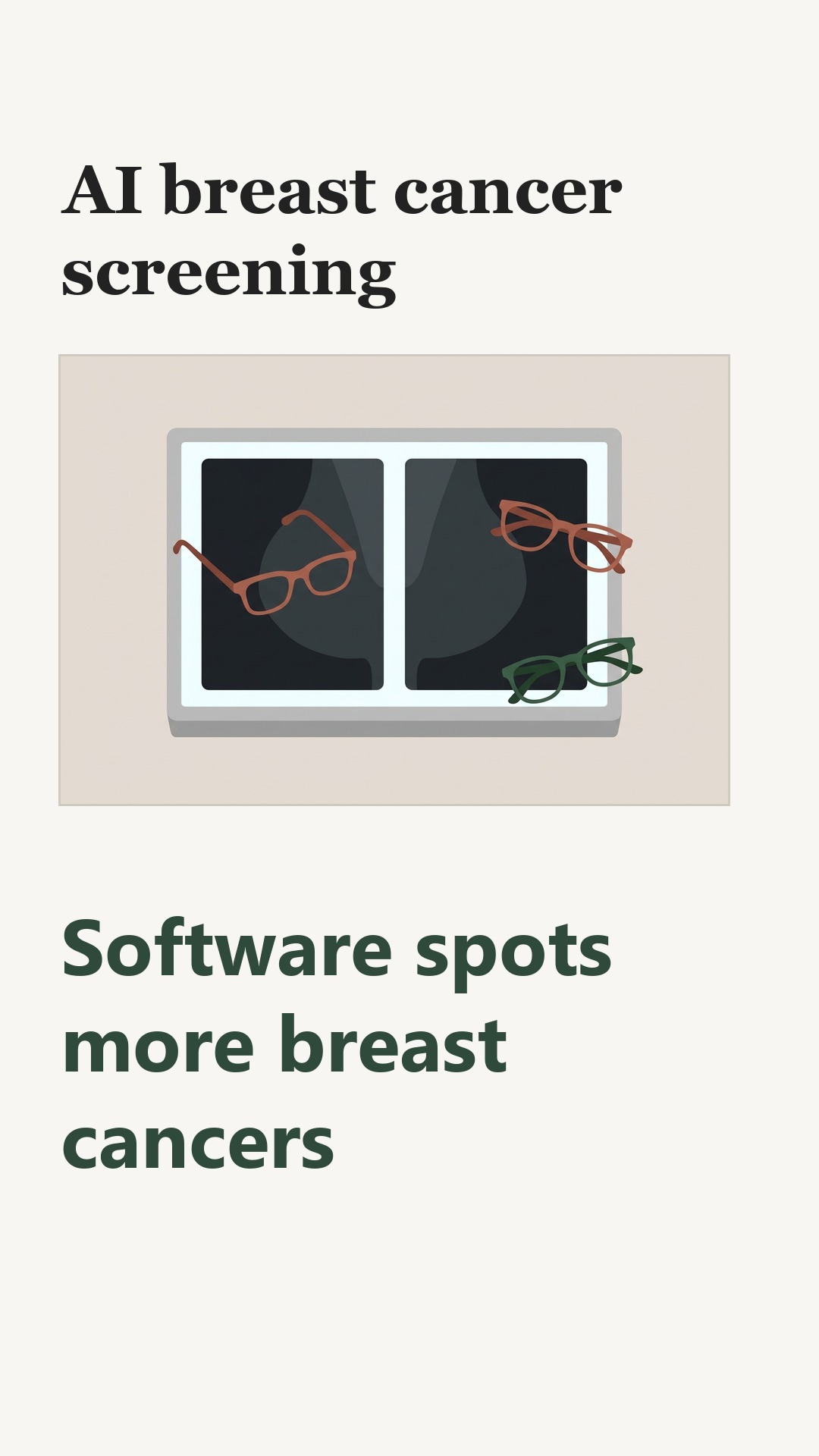
In Sweden's national breast screening program, a computer program was given a job. It read each woman's mammogram and gave it a risk score.
The article describes a trial that enrolled 105,934 women and assigned them at random, half to the usual reading and half to AI-supported reading.
Consider what the program did with the score. The article describes a trial in which low-risk mammograms went to one radiologist instead of two, while higher-risk ones still went to two. The article describes a trial in which radiologists read 44.3 percent fewer screens that way, a saving any understaffed clinic would notice.
The article describes a trial in which the scans read with the software's help turned up 29 percent more cancers, 6.4 per 1,000 women against 5.0. The article describes a trial in which most of these cancers were small and had not reached the lymph nodes. The article describes a trial in which cancers found between scheduled screenings fell 12 percent.
The article describes a trial in which the share of women wrongly flagged did not go up. The article describes a trial in which the share of women correctly cleared was 98.5 percent in both arms.
Grant the trial this much: it measured what screening found, not what happened to the women afterward. The article says that no completed study of AI screening has shown fewer breast cancer deaths. More cancers found is not the same as longer life.
Some of the extra finds may be slow-growing cancers that would never have harmed anyone. The article describes a trial that found more of the low-grade and in-situ kinds, which can lead to biopsy and treatment a woman did not need. The article says later screening rounds will help sort this out.
The women were screened in Sweden, in a program that normally uses two readers. A clinic that uses one reader, or screens a different population, cannot assume the same results.
So the question to carry into your own clinic is not whether the software works. It is whether the program near you uses such a tool, and whether it was tested on people like you.
What this means for you
When a clinic near you adopts a tool like this, the fair question is whether it serves women like you and whether anyone followed them afterward. In Sweden the program still used two readers, so a place that reads once cannot simply borrow the result. Ask your clinic what it uses and who tested it.
Kumar, A., Lip, G. (2026). Artificial Intelligence in Breast Imaging and Screening: Current Evidence, Applications, and Future Directions. Indian Journal of Radiology and Imaging. https://doi.org/10.1055/s-0046-1827249
Who paid: The article states no funding source and declares no conflicts of interest; it does not say who funded the study or lent equipment or software.
Do not take this as professional medical advice.
The findings of other studies mentioned here are known to us through this document, which is the one we read; we did not open each of those studies.
The longer versionThe passages copied from the paper, the pictures, and every source with how much of it we read · 1384 words · about 7 minRead it →Close
AI Helped Swedish Mammograms Find More Cancers. No Trial Yet Shows Fewer Deaths.
What the largest randomized test of screening software found, what it did not, and what to ask before your clinic installs one.
Consider the mammogram a woman in Sweden gets every two years. Two radiologists read it, one after the other, because two pairs of eyes catch more cancers than one. That second read is also why a screening program needs so many radiologists, and why a global shortage of breast radiologists makes the second read harder to sustain1.
The Mammography Screening with Artificial Intelligence trial, or MASAI, took a different approach. Software gave every mammogram a risk score. Low-risk cases went to a single radiologist instead of two. Higher-risk cases kept the standard two-reader review2. The trial enrolled 105,934 women in the Swedish national screening program, randomized in equal halves to AI-supported screening or standard double reading.
The results arrived in three stages. The article describes a study in which the 2023 safety analysis found 20 percent more cancers detected — 6.1 versus 5.1 per 1,000 women — with no change in the false-positive rate, and 44.3 percent less screen-reading work for radiologists3. The article describes a study in which the 2025 performance analysis put the detection gain at 29 percent (6.4 versus 5.0 per 1,000), mostly small, lymph-node-negative cancers, including 27 percent more aggressive subtypes4. The article describes a study in which the 2026 primary endpoint found higher sensitivity, 80.5 percent versus 73.8 percent, with identical specificity of 98.5 percent, and a 12 percent lower interval cancer rate — cancers that surface between screenings5.
Picture the workload. A 44.3 percent cut means a department that needed roughly nine radiologist-hours to read a batch of screening mammograms now needs about five3. That is not a rounding error; it is the difference between sustaining double reading and abandoning it.
The Swedish trial is not the only evidence, and the other studies matter because they tested different setups. The article describes a study in which a German program evaluated AI-supported double reading in more than 460,000 women and reported a 17.6 percent detection increase (6.7 versus 5.7 per 1,000) without raising the recall rate (37.4 versus 38.3 per 1,000)6. The article describes a study in which a Spanish study of 31,301 women let the software mark low-risk cases as normal without any radiologist reading them; workload fell 63.6 percent and detection rose 15.2 percent7. The article describes a study in which a South Korean study of 24,543 women tested the software in a single-read system and found detection at 5.70 versus 5.01 per 1,000, with no significant difference in recalls8. A United Kingdom study, GEMINI, tested 17 different ways of fitting the software into a screening program and found that some setups improved every measured metric, with workload savings of up to 36 percent.
The article describes a study in which a systematic review of 31 studies covering more than two million screening examinations found that triage setups — the kind MASAI used — delivered the largest operational benefit, cutting reading volumes by 40 to 90 percent while cancer detection stayed noninferior9.
The limits are as important as the numbers. The review that gathered this evidence notes that the evidence comes mostly from non-diverse, high-income populations, and that long-term data including breast cancer mortality are not yet available from any completed AI screening study10. The article describes a study in which the MASAI trial found more low-grade luminal A cancers and more in-situ lesions alongside the aggressive cancers it caught — a pattern the authors say warrants scrutiny for overdiagnosis, the detection of slow-growing disease that would never have harmed anyone. And the Swedish result belongs to one configuration inside one country's double-reading program: a partial substitution, not a replacement of radiologists2.
Consider what the software actually does, because the difference between setups decides what a reader gets. In a second-reader setup, the software reviews the image after a radiologist and offers a second opinion. In a triage setup, it sorts cases by risk so that only the higher-risk ones get two readers. In a partially autonomous setup, it clears low-risk cases with no radiologist involved at all2. The last one is the most efficient and the least governed: the European Society of Breast Imaging recommends that AI findings stay subject to radiologist oversight and be overrulable.
The evidence has a geography problem. The article describes a study in which a review of the research found that only 0 to 25 percent of studies reported patients' race or ethnicity, most patients were identified as Caucasian, and nearly all cohorts came from high-income countries, with no studies from low-income settings11. The article describes a study in which researchers applied models trained on large but less diverse data to a heterogeneous California population, and individual models reached sensitivity of only 0.4 to 0.512. A separate review of the field, which we could read only in its summary because the full paper sits behind a subscription, concluded that pooled accuracy is high on average but so dispersed that it cannot be transferred to a new program, and that local validation before deployment remains necessary13.
Here is how we read it. Software learns from the images it is shown, and it tends to do best on images that resemble them. A screening program in Latin America, or one serving patients unlike the Swedish and British women in these trials, may find the advantage smaller or shifted, and may need to adjust its thresholds before trusting the extra cancers found. We would expect that adjustment to be needed, not optional. You would know we are wrong if a diverse public program ran its own pilot and reported the same detection gain and workload cut with no retuning. When a clinic announces AI-assisted mammography, ask whether the tool was tested on patients like the local population, and whether results are being tracked after rollout.
There is a quieter risk. People tend to accept a confident automated answer at face value, especially when it says everything is fine, and to stop checking as carefully themselves. In a program where the software clears most mammograms for a single reader, that lone reader may grow less scrutinizing over months, and a cancer the software missed could slip through. Ask whether your program shows the software's result only after a radiologist has read the image, and whether it audits how often readers overrule it.
A tool can genuinely help and still be sold with claims that outrun what was measured. We would expect clinics to advertise "more cancers found" without mentioning that no trial has yet shown fewer deaths, and without saying whether the extra finds were harmful or harmless. You would know we are wrong if programs published their own before-and-after figures, including deaths and overdiagnosis, alongside detection numbers. When you hear an AI screening claim, ask what was measured, in how many people, and over how long.
The regulatory picture differs by country, and that shapes what reaches a clinic near you. In the United States, most breast-imaging AI tools enter through a premarket notification pathway based on similarity to an existing device. In the European Union, they are regulated as medical devices with additional transparency and human-oversight requirements for high-risk uses including cancer screening. The review's authors call for rigorous post-market surveillance, inclusive datasets and cost-effectiveness analyses before the field moves to routine use14.
Other trials are under way in Norway, Australia and the United Kingdom, and their long-term results will speak to the question MASAI cannot yet answer. A separate review of the past decade of this research, which we could read only in its summary, put it this way: the evidence supports AI as a second reader and triage tool in organized screening, not as an autonomous replacement for the radiologist15.
The mammogram still gets read by a person in every setup tested so far. What changed in Sweden is how many people read it, and which ones. If your screening program adopts software like this, the useful question is not whether it is impressive but whether anyone checked it on people like you — and whether anyone is still counting the cancers it misses.
What would you ask your screening clinic before trusting an AI-assisted mammogram?
Where each piece of context comes from, and how much of it we read
- Kumar, A., Lip, G. (2026). Artificial Intelligence in Breast Imaging and Screening: Current Evidence, Applications, and Future Directions. Indian Journal of Radiology and Imaging. https://doi.org/10.1055/s-0046-1827249 - the article this story is about — the whole article — the passage: "European guidelines recommend double reading of screening mammograms to ensure high sensitivity, but this practice imposes a substantial workload burden and is increasingly difficult to sustain due to a global shortage of breast radiologists."
- Kumar, A., Lip, G. (2026). Artificial Intelligence in Breast Imaging and Screening: Current Evidence, Applications, and Future Directions. Indian Journal of Radiology and Imaging. https://doi.org/10.1055/s-0046-1827249 - the article this story is about — the whole article — the passage: "Contemporary DL systems can be deployed in several operational configurations: as standalone AI (operating independently of a radiologist); as second-reader AI (reviewing cases after an initial radiologist read and functioning as a second opinion); within triage workflows (stratifying cases by risk level to direct reading resource, with low-risk cases directed to single reading and higher risk cases to standard double reading); or in partially autonomous workflows (where AI classifies low-risk cases as normal without radiologist review, reserving reading resource for AI-flagged cases only)."
- Kumar, A., Lip, G. (2026). Artificial Intelligence in Breast Imaging and Screening: Current Evidence, Applications, and Future Directions. Indian Journal of Radiology and Imaging. https://doi.org/10.1055/s-0046-1827249 - the article this story is about — the whole article — the passage: "The initial safety analysis (2023) demonstrated that AI-supported screening detected 20% more cancers (cancer detection rate [CDR] 6.1 vs. 5.1 per 1,000; above the lowest acceptable safety limit) without affecting the false-positive rate, while achieving a 44.3% reduction in screen-reading workload."
- Kumar, A., Lip, G. (2026). Artificial Intelligence in Breast Imaging and Screening: Current Evidence, Applications, and Future Directions. Indian Journal of Radiology and Imaging. https://doi.org/10.1055/s-0046-1827249 - the article this story is about — the whole article — the passage: "A subsequent analysis of screening performance (2025) confirmed a 29% increase in cancer detection (CDR 6.4 vs. 5.0 per 1,000; p = 0.0021), predominantly of small, lymph-node-negative invasive cancers, including 27% more aggressive nonluminal A subtypes (triple-negative, HER2-positive, and luminal B)."
- Kumar, A., Lip, G. (2026). Artificial Intelligence in Breast Imaging and Screening: Current Evidence, Applications, and Future Directions. Indian Journal of Radiology and Imaging. https://doi.org/10.1055/s-0046-1827249 - the article this story is about — the whole article — the passage: "AI-supported screening achieved higher sensitivity (80.5% vs. 73.8%) with identical specificity (98.5%) compared with standard double reading. The interval cancer rate was reduced by 12%, meeting the criterion for noninferiority."
- Kumar, A., Lip, G. (2026). Artificial Intelligence in Breast Imaging and Screening: Current Evidence, Applications, and Future Directions. Indian Journal of Radiology and Imaging. https://doi.org/10.1055/s-0046-1827249 - the article this story is about — the whole article — the passage: "The study found that AI-supported double reading increased the breast cancer detection rate by 17.6% (6.7 vs. 5.7 per 1,000 women) compared to standard double reading, without increasing the recall rate (37.4 vs. 38.3 per 1,000)."
- Kumar, A., Lip, G. (2026). Artificial Intelligence in Breast Imaging and Screening: Current Evidence, Applications, and Future Directions. Indian Journal of Radiology and Imaging. https://doi.org/10.1055/s-0046-1827249 - the article this story is about — the whole article — the passage: "Radiologist workload was reduced by 63.6%, and the CDR increased by 15.2% (from 6.3 to 7.3 per 1,000, p < 0.001)."
- Kumar, A., Lip, G. (2026). Artificial Intelligence in Breast Imaging and Screening: Current Evidence, Applications, and Future Directions. Indian Journal of Radiology and Imaging. https://doi.org/10.1055/s-0046-1827249 - the article this story is about — the whole article — the passage: "The CDR was significantly higher with AI-CAD (5.70‰ vs. 5.01‰, p < 0.001) with no significant difference in recall rates."
- Kumar, A., Lip, G. (2026). Artificial Intelligence in Breast Imaging and Screening: Current Evidence, Applications, and Future Directions. Indian Journal of Radiology and Imaging. https://doi.org/10.1055/s-0046-1827249 - the article this story is about — the whole article — the passage: "When used as a second reader or within double-reading workflows, AI generally maintained or modestly increased sensitivity (up to +9 percentage points) while preserving or improving specificity. Triage configurations delivered the greatest operational benefit, reducing reading volumes by 40 to 90% while maintaining noninferior cancer detection."
- Kumar, A., Lip, G. (2026). Artificial Intelligence in Breast Imaging and Screening: Current Evidence, Applications, and Future Directions. Indian Journal of Radiology and Imaging. https://doi.org/10.1055/s-0046-1827249 - the article this story is about — the whole article — the passage: "However, the evidence base remains predominantly derived from nondiverse, high-income country populations, and long-term outcome data including breast cancer mortality are not yet available."
- Kumar, A., Lip, G. (2026). Artificial Intelligence in Breast Imaging and Screening: Current Evidence, Applications, and Future Directions. Indian Journal of Radiology and Imaging. https://doi.org/10.1055/s-0046-1827249 - the article this story is about — the whole article — the passage: "Only 0 to 25% of studies reported race/ethnicity, with most patients identified as Caucasian. Nearly all patient cohorts originated from high-income countries, with no studies from low-income settings."
- Kumar, A., Lip, G. (2026). Artificial Intelligence in Breast Imaging and Screening: Current Evidence, Applications, and Future Directions. Indian Journal of Radiology and Imaging. https://doi.org/10.1055/s-0046-1827249 - the article this story is about — the whole article — the passage: "Hsu et al demonstrated that AI models trained on large but less diverse datasets substantially underperformed when applied to the heterogeneous University of California Los Angeles (UCLA) population, with individual models achieving sensitivity of only 0.4 to 0.5."
- Ciurescu S, Buciu V, Ilaș DG, Pârvănescu R, Șerban D. (2026). Ten Years of Artificial Intelligence in Screening Mammography: A Systematic Review and Meta-Analysis of Diagnostic Accuracy and Clinical Implementation (Literature Published 2015–2025). Diagnostics. 10.3390/diagnostics16183045 — only the abstract - the full text could not be fetched — the passage: "Pooled accuracy is high on average but so dispersed that it cannot be transferred to a new programme; local validation before deployment remains necessary."
- Kumar, A., Lip, G. (2026). Artificial Intelligence in Breast Imaging and Screening: Current Evidence, Applications, and Future Directions. Indian Journal of Radiology and Imaging. https://doi.org/10.1055/s-0046-1827249 - the article this story is about — the whole article — the passage: "As the field transitions from research to implementation, rigorous post-market surveillance, inclusive dataset development, and cost-effectiveness analyses will be essential to ensure that AI delivers equitable improvements in breast cancer care."
- Ciurescu S, Buciu V, Ilaș DG, Pârvănescu R, Șerban D. (2026). Ten Years of Artificial Intelligence in Screening Mammography: A Systematic Review and Meta-Analysis of Diagnostic Accuracy and Clinical Implementation (Literature Published 2015–2025). Diagnostics. 10.3390/diagnostics16183045 — only the abstract - the full text could not be fetched — the passage: "A decade of evidence supports AI as a second reader and triage tool in organised screening, not as an autonomous replacement for the radiologist."
Kumar, A., Lip, G. (2026). Artificial Intelligence in Breast Imaging and Screening: Current Evidence, Applications, and Future Directions. Indian Journal of Radiology and Imaging. https://doi.org/10.1055/s-0046-1827249
Who paid: The article states no funding source and declares no conflicts of interest; it does not say who funded the study or lent equipment or software.
Do not take this as professional medical advice.
The findings of other studies mentioned here are known to us through this document, which is the one we read; we did not open each of those studies.