other · Gastroenterology report · la publicación, 12 sep 2026 · gratis
La IA ayudó a los médicos a mirar más y mejor un órgano difícil de ver
Un estudio en un solo hospital de China halló más exámenes completos y más masas pequeñas de páncreas. No prueba que los pacientes vivan más, y nadie debe saltarse un examen por esto.
Versión breve · la versión detallada sigue, unos 6 min
- El estudio, de un vistazo
- Quiénes
- Exámenes de ultrasonido endoscópico de páncreas y vías biliares
- Cuántos
- 854 exámenes, 427 en cada grupo
- Dónde
- Hospital Renmin de la Universidad de Wuhan, China
- Cuándo
- Entre noviembre de 2016 y diciembre de 2025
- Tipo de estudio
- análisis de lo que hicieron los médicos en dos épocas distintas
- Quién lo hizo
- Hospital Renmin de la Universidad de Wuhan, China
- El límite que importa
- Un solo hospital, mirada hacia atrás: muestra asociación, no prueba causa
Porcentaje de estaciones anatómicas documentadas y de exámenes donde se detectaron masas de páncreas menores de 2 cm, antes y después de incorporar el sistema de IA. Son dos medidas distintas, no una sola; el estudio fue observacional en un solo hospital.
Exámenes antes de la IA frente a exámenes después de la IA
Se documentaron más estaciones anatómicas: 89.93% frente a 81.29%
Se detectaron más masas de páncreas menores de 2 cm: 11.48% frente a 6.56%
Se detectaron más lesiones biliopancreáticas en total: 44.50% frente a 36.53%
Se detectaron más masas en cuerpo y cola del páncreas: 13.11% frente a 8.43%
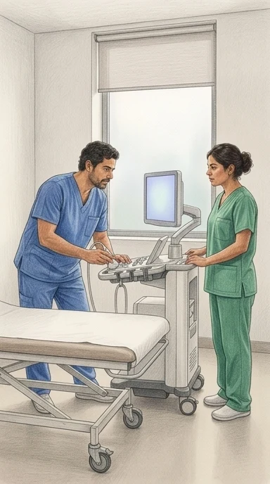
Para revisar el páncreas y las vías biliares, el médico introduce una sonda por la boca y usa el ultrasonido desde adentro. Es una de las pruebas más difíciles de la endoscopia: hay que mover el aparato, orientarse en el espacio y reconocer estructuras a la vez. Si el especialista no llega a una zona, esa zona simplemente no se vio.
Ocho puntos son los que se deben fotografiar para dejar constancia de que el recorrido quedó completo. Antes de la inteligencia artificial, en este hospital solo se documentaba alrededor del 81 por ciento de esos ocho puntos.
Investigadores del Hospital Renmin de la Universidad de Wuhan, en China, compararon 427 exámenes hechos antes de incorporar el sistema y 427 después, igualados por edad, sexo, origen del paciente y experiencia del médico. El sistema, llamado EUS-IREAD, muestra en una pantalla secundaria qué zonas ya se recorrieron y cuáles faltan, mientras el examen ocurre.
Con el sistema, la constancia subió a cerca del 90 por ciento de los puntos. La detección de masas de páncreas menores de 2 centímetros pasó de 6.56 a 11.48 por ciento. La mejora mayor se dio en la cola del páncreas y en el hilio del hígado, dos zonas esquivas.
Conviene ser claro: fue un solo hospital, mirando hacia atrás en los registros, no un ensayo aleatorizado en su comunidad. Muestra una asociación, no prueba que la IA cause mejores resultados. También conviene notar algo que el estudio no demuestra: no aumentaron los cánceres de páncreas detectados, ni en total ni entre los menores de 2 centímetros. Muchas de esas masas pequeñas pueden no ser malignas; el hallazgo no equivale todavía a un diagnóstico más temprano ni a más años de vida. Tampoco se midió si los pacientes estuvieron mejor después, y el seguimiento no fue parejo.
Los médicos con más de 2,000 procedimientos fueron quienes más mejoraron su constancia. Los autores sugieren que esto podría deberse a que suelen adoptar una estrategia de examen más dirigida, basada en su sospecha clínica previa y su experiencia, lo que puede reducir la constancia en la documentación de los puntos anatómicos cuando no hay una enfermedad evidente.
Los autores señalan que este tipo de sistema actúa antes de que aparezca una lesión: ayuda a que el examen cubra lo que debe cubrir. Si alguna vez le piden un ultrasonido endoscópico de páncreas o vías biliares, pregunte si el centro donde lo harán usa protocolos de puntos anatómicos definidos y cómo verifican que el recorrido quedó completo. Nunca aplace ni cancele un examen por esperar una tecnología así.
Qué significa para usted
Si le piden un ultrasonido endoscópico de páncreas o vías biliares, pregunte en el centro si usan protocolos con puntos anatómicos definidos y cómo comprueban que el recorrido quedó completo. Sepa que esta ayuda mostró exámenes más completos y más masas pequeñas detectadas en un solo hospital de China, pero no probó que alguien viva más ni que se detecte antes el cáncer. Nunca aplace ni cancele un examen por esperar esa tecnología.
Zhang, C., Zhang, J., Tan, W. et al. (2026). Real-time AI assistance improves adherence to standardized biliopancreatic EUS examination: a real-world comparative study. Gastroenterology Report. https://doi.org/10.1093/gastro/goag078
Quién pagó: El estudio fue financiado por el Programa Clave de Investigación y Desarrollo de la Provincia de Hubei (No. 2023BCB153), el Proyecto del Centro Provincial de Investigación Clínica de Hubei para Incisión Mínimamente Invasiva en Enfermedades Digestivas (No. 2024CCB007), el Proyecto de Escenario de Demostración de Aplicación de Inteligencia Artificial de Wuhan (No. 2022YYCJ01) y el Proyecto de Reforma Educativa de la Facultad de Medicina de la Universidad de Wuhan (No. 2024YB28); el artículo no indica que los financiadores tuvieran participación alguna en el diseño, análisis o reporte, y no se mencionan préstamos de equipos o software.
No tome esto como consejo médico profesional.
Versión detalladaLos pasajes copiados del artículo, las ilustraciones y cada fuente con cuánto leímos de ella · 1116 palabras · unos 6 minLeerla →Cerrar
La IA ya no solo busca lesiones: ahora vigila que el médico no se salte ninguna zona del examen
Un estudio retrospectivo en un hospital chino encontró que un sistema de ayuda en tiempo real se asoció con más estaciones anatómicas documentadas y más masas pequeñas de páncreas detectadas. No probó que los pacientes vivan más.
El ultrasonido endoscópico del páncreas y las vías biliares es uno de los exámenes más difíciles de hacer bien en toda la endoscopia digestiva. Quien lo realiza debe manejar el endoscopio, orientarse en el espacio, interpretar lo que ve y decidir en el momento, todo a la vez1. Por eso quedan zonas sin revisar y los informes varían de un médico a otro2. Ese es el problema que un grupo del Hospital Renmin de la Universidad de Wuhan, en China, quiso atacar con un sistema de inteligencia artificial llamado EUS-IREAD, que ya habían probado antes en un ensayo controlado.
El estudio que ahora publicaron no es un experimento nuevo: es una mirada hacia atrás a los registros de 1,465 exámenes hechos entre noviembre de 2016 y diciembre de 2025. Compararon los exámenes hechos antes de que el sistema entrara en la rutina diaria con los hechos después, dejando fuera un período intermedio de casi dos años para no mezclar el efecto de la instalación. Después de igualar los dos grupos por edad, sexo, tipo de paciente y experiencia del endoscopista, quedaron 854 exámenes, 427 en cada lado. Es un estudio observacional: no se sortearon los pacientes, se compararon dos épocas.
¿Qué encontraron? Que en la etapa con ayuda de la IA se documentaron más de las ocho estaciones anatómicas que el protocolo exige fotografiar: 89.93% de las estaciones en promedio, frente a 81.29% antes3. Y que se detectaron más masas de páncreas menores de 2 centímetros: 11.48% de los exámenes frente a 6.56%4. También subió la detección de lesiones biliopancreáticas en conjunto5 y de masas en cuerpo y cola del páncreas6. Cuando repitieron el análisis restringiéndose a una ventana simétrica de 48 meses alrededor del período excluido, los resultados se mantuvieron7.
Pero hay un dato que obliga a bajarle el tono a la noticia: no aumentó la detección de cáncer de páncreas, ni en total ni entre los tumores menores de 2 centímetros8. Es decir, se encontraron más masas pequeñas, pero no un aumento claro de cánceres. No todas las masas pequeñas son malignas: algunas son inflamatorias, benignas o indeterminadas. Además, como el estudio es retrospectivo, el seguimiento de esos pacientes no fue uniforme y no se sabe bien qué pasó después con ellos; harían falta estudios prospectivos con seguimiento estandarizado para saber si esas lesiones adicionales cambiaron algo en la vida de alguien9. El estudio se hizo en un solo hospital, con diseño histórico y no aleatorio, así que no se puede descartar que otros cambios del período expliquen parte del resultado10. Y fue en un centro terciario con mucha experiencia en este tipo de examen, lo que limita generalizar a otros lugares11.
Para entender qué se probó, hay que entender el mecanismo. El sistema no busca lesiones en la imagen como otros programas de inteligencia artificial en medicina. Lo que hace es mostrar en una pantalla secundaria, mientras el médico trabaja, un mapa de las estaciones anatómicas ya revisadas y las que faltan, además de tiempos, avisos de estructuras requeridas, ayuda para segmentar imágenes y medición automática de conductos12. La función que les interesaba medir era la de apoyo a nivel de estaciones: indicar visualmente qué zonas del recorrido ya se escanearon y cuáles no, para que el operador complete el camino estandarizado13. Es, en términos simples, un copiloto del recorrido, no un detector de tumores.
El problema de fondo es conocido. En endoscopia digestiva, la mayoría de los sistemas de IA se han dedicado a detectar o diagnosticar lesiones una vez que la lesión ya está en el campo de visión. Pero en este examen el resultado depende de algo anterior: que la zona donde está la lesión se haya mirado. Si una estación no se explora, ningún algoritmo puede compensarlo. Y en lugares con mucho volumen de trabajo, cumplir el protocolo completo compite con la eficiencia diaria14. Ahí es donde entra la propuesta de este grupo.
Hay una comparación que el propio estudio permite hacer y que da contexto al hallazgo. Antes de la IA, los endoscopistas expertos (más de 2,000 procedimientos) documentaban menos estaciones que los de menor experiencia; después de la IA, los expertos mejoraron más que los demás, con un aumento de 13,4 puntos frente a 7,215. Es decir, el sistema pareció ayudar sobre todo a quienes más se saltaban pasos por criterio propio, no a los que recién aprenden. El estudio mismo advierte que esta parte debe tomarse como exploratoria, porque la distribución de endoscopistas entre los dos períodos era muy distinta.
Así lo leemos nosotros. Lo que este trabajo sugiere es un cambio de conversación: la IA en medicina no solo puede servir para reconocer algo en una imagen, sino para vigilar que el procedimiento se haga completo. Es una idea distinta y menos vistosa. Si eso fuera cierto en la práctica, lo que usted podría esperar es que en un centro que use una guía así, el informe mencione de forma sistemática todas las zonas recomendadas, incluidas las más difíciles de alcanzar, como la cola del páncreas. Sabríamos que nos equivocamos si en otros hospitales, con otros médicos y otros pacientes, la diferencia desapareciera, o si al retirar el sistema las cifras volvieran a bajar. Por ahora, lo que hay es una asociación medida en un solo centro, no una promesa.
Hay algo más que conviene tener presente. Cuando se introduce una herramienta que promete mejorar y todos saben que se está evaluando, parte del cambio puede venir de sentirse observado, no de la herramienta. Este estudio no comparó contra un grupo igualmente vigilado pero sin IA, así que esa duda queda abierta.
¿Qué puede hacer usted con esto? Si a usted o a un familiar le hacen un ultrasonido endoscópico del páncreas o las vías biliares, puede preguntar si el centro sigue un protocolo fijo de estaciones anatómicas y si el informe las documenta todas. Puede preguntar si el médico adapta el examen según lo que sospecha o lo completa igual. Y puede pedir que le expliquen qué zonas se revisaron y cuáles no. No hay que cambiar ningún tratamiento por esto, ni retrasar nada: es una pregunta más para llevar a la consulta. La tecnología todavía no demostró que alargue vidas, pero sí demostró, en este hospital, que ayuda a que el examen se haga completo. ¿Sabe usted si el centro donde se atiende documenta las ocho estaciones?
De dónde sale cada dato de contexto, y cuánto leímos de cada documento
- Zhang, C., Zhang, J., Tan, W. et al. (2026). Real-time AI assistance improves adherence to standardized biliopancreatic EUS examination: a real-world comparative study. Gastroenterology Report. https://doi.org/10.1093/gastro/goag078 - el artículo del que trata esta nota — el artículo completo — el pasaje: "EUS remains one of the most operator-dependent procedures in gastrointestinal endoscopy because it requires simultaneous scope manipulation, spatial orientation, anatomical interpretation, and real-time clinical decision-making"
- Zhang, C., Zhang, J., Tan, W. et al. (2026). Real-time AI assistance improves adherence to standardized biliopancreatic EUS examination: a real-world comparative study. Gastroenterology Report. https://doi.org/10.1093/gastro/goag078 - el artículo del que trata esta nota — el artículo completo — el pasaje: "As a result, incomplete examination of key anatomical regions and inconsistent procedural documentation remain important challenges in routine practice"
- Zhang, C., Zhang, J., Tan, W. et al. (2026). Real-time AI assistance improves adherence to standardized biliopancreatic EUS examination: a real-world comparative study. Gastroenterology Report. https://doi.org/10.1093/gastro/goag078 - el artículo del que trata esta nota — el artículo completo — el pasaje: "Adherence to the standardized eight-station examination was higher in the post-AI group, reflected by greater total documentation completeness (89.93% vs 81.29%, adjusted absolute difference 8.65%, 95% confidence interval [CI] 6.58–10.71, P < 0.001)."
- Zhang, C., Zhang, J., Tan, W. et al. (2026). Real-time AI assistance improves adherence to standardized biliopancreatic EUS examination: a real-world comparative study. Gastroenterology Report. https://doi.org/10.1093/gastro/goag078 - el artículo del que trata esta nota — el artículo completo — el pasaje: "Detection of pancreatic masses smaller than 2 cm was also higher after AI implementation (11.48% vs 6.56%, adjusted odds ratio 1.82, 95% CI 1.13–3.01, P = 0.016)."
- Zhang, C., Zhang, J., Tan, W. et al. (2026). Real-time AI assistance improves adherence to standardized biliopancreatic EUS examination: a real-world comparative study. Gastroenterology Report. https://doi.org/10.1093/gastro/goag078 - el artículo del que trata esta nota — el artículo completo — el pasaje: "In the matched cohort, the post-AI group showed a higher detection rate of total biliopancreatic lesions than the pre-AI group (44.50% vs 36.53%, aOR 1.40, 95% CI 1.04–1.87, P = 0.026)."
- Zhang, C., Zhang, J., Tan, W. et al. (2026). Real-time AI assistance improves adherence to standardized biliopancreatic EUS examination: a real-world comparative study. Gastroenterology Report. https://doi.org/10.1093/gastro/goag078 - el artículo del que trata esta nota — el artículo completo — el pasaje: "Detection of body/tail pancreatic masses was also higher after AI implementation (13.11% vs 8.43%, aOR 1.62, 95% CI 1.04–2.55, P = 0.036)."
- Zhang, C., Zhang, J., Tan, W. et al. (2026). Real-time AI assistance improves adherence to standardized biliopancreatic EUS examination: a real-world comparative study. Gastroenterology Report. https://doi.org/10.1093/gastro/goag078 - el artículo del que trata esta nota — el artículo completo — el pasaje: "In the sensitivity analysis restricted to a symmetrical 48-month window surrounding the washout period, the primary findings remained consistent."
- Zhang, C., Zhang, J., Tan, W. et al. (2026). Real-time AI assistance improves adherence to standardized biliopancreatic EUS examination: a real-world comparative study. Gastroenterology Report. https://doi.org/10.1093/gastro/goag078 - el artículo del que trata esta nota — el artículo completo — el pasaje: "Although pancreatic masses smaller than 2 cm were detected more frequently after AI implementation, this signal was not accompanied by a significant increase in pancreatic cancer detection, either overall or among lesions smaller than 2 cm."
- Zhang, C., Zhang, J., Tan, W. et al. (2026). Real-time AI assistance improves adherence to standardized biliopancreatic EUS examination: a real-world comparative study. Gastroenterology Report. https://doi.org/10.1093/gastro/goag078 - el artículo del que trata esta nota — el artículo completo — el pasaje: "Because final lesion classification relied on pathological findings when available and available clinical follow-up, and because follow-up duration was not uniform, prospective studies with standardized follow-up are needed to determine whether the additional small lesions identified after AI implementation are clinically actionable."
- Zhang, C., Zhang, J., Tan, W. et al. (2026). Real-time AI assistance improves adherence to standardized biliopancreatic EUS examination: a real-world comparative study. Gastroenterology Report. https://doi.org/10.1093/gastro/goag078 - el artículo del que trata esta nota — el artículo completo — el pasaje: "First, this was a retrospective single-center study with a historical control design, and residual confounding cannot be excluded despite the use of PSM and ITS."
- Zhang, C., Zhang, J., Tan, W. et al. (2026). Real-time AI assistance improves adherence to standardized biliopancreatic EUS examination: a real-world comparative study. Gastroenterology Report. https://doi.org/10.1093/gastro/goag078 - el artículo del que trata esta nota — el artículo completo — el pasaje: "Fifth, the study was conducted at a tertiary referral center with substantial expertise in biliopancreatic EUS, which may limit generalizability to other practice settings."
- Zhang, C., Zhang, J., Tan, W. et al. (2026). Real-time AI assistance improves adherence to standardized biliopancreatic EUS examination: a real-world comparative study. Gastroenterology Report. https://doi.org/10.1093/gastro/goag078 - el artículo del que trata esta nota — el artículo completo — el pasaje: "During the AI-assisted phase, the system provided real-time workflow feedback through a secondary monitor, including procedure timing, anatomical station monitoring, prompts for required anatomical structures, real-time segmentation assistance, and automated duct measurement."
- Zhang, C., Zhang, J., Tan, W. et al. (2026). Real-time AI assistance improves adherence to standardized biliopancreatic EUS examination: a real-world comparative study. Gastroenterology Report. https://doi.org/10.1093/gastro/goag078 - el artículo del que trata esta nota — el artículo completo — el pasaje: "For the present study, the function of primary interest was station-level examination support, which visually indicated scanned and unscanned predefined biliopancreatic stations to assist operators in completing the standardized examination pathway."
- Zhang, C., Zhang, J., Tan, W. et al. (2026). Real-time AI assistance improves adherence to standardized biliopancreatic EUS examination: a real-world comparative study. Gastroenterology Report. https://doi.org/10.1093/gastro/goag078 - el artículo del que trata esta nota — el artículo completo — el pasaje: "In daily clinical work, however, adherence to such protocols may be suboptimal, particularly in high-volume settings where endoscopists must balance procedural efficiency with diagnostic thoroughness"
- Zhang, C., Zhang, J., Tan, W. et al. (2026). Real-time AI assistance improves adherence to standardized biliopancreatic EUS examination: a real-world comparative study. Gastroenterology Report. https://doi.org/10.1093/gastro/goag078 - el artículo del que trata esta nota — el artículo completo — el pasaje: "Before AI implementation, expert endoscopists showed lower documentation completeness than senior endoscopists, whereas after implementation, the absolute increase in total documentation completeness was greater among experts than among seniors (13.4% vs. 7.2%, P for interaction <0.001)"
Zhang, C., Zhang, J., Tan, W. et al. (2026). Real-time AI assistance improves adherence to standardized biliopancreatic EUS examination: a real-world comparative study. Gastroenterology Report. https://doi.org/10.1093/gastro/goag078
Quién pagó: El estudio fue financiado por el Programa Clave de Investigación y Desarrollo de la Provincia de Hubei (No. 2023BCB153), el Proyecto del Centro Provincial de Investigación Clínica de Hubei para Incisión Mínimamente Invasiva en Enfermedades Digestivas (No. 2024CCB007), el Proyecto de Escenario de Demostración de Aplicación de Inteligencia Artificial de Wuhan (No. 2022YYCJ01) y el Proyecto de Reforma Educativa de la Facultad de Medicina de la Universidad de Wuhan (No. 2024YB28); el artículo no indica que los financiadores tuvieran participación alguna en el diseño, análisis o reporte, y no se mencionan préstamos de equipos o software.
No tome esto como consejo médico profesional.
other · Gastroenterology report · the paper, 12 Sep 2026 · free
AI Guidance Helped Doctors Cover More Ground in Pancreas Exams
One hospital's records show more complete scans and more small masses found — but not more cancers, and not proof that patients fared better.
Short version · the longer version follows, about 6 min
- The study at a glance
- Who
- people who had an ultrasound exam of the pancreas and bile ducts
- How many
- 854 matched examinations (427 per group)
- Where
- Renmin Hospital of Wuhan University, China
- When
- examinations from 1 November 2016 to 17 December 2025
- Kind of study
- analysis of what people did (a look back at records)
- Who did it
- Renmin Hospital of Wuhan University, China
- The limit that matters
- One hospital, old records, no proof the AI caused the results.
These are the shares of the eight required stations that were photographed during each exam, in one hospital's records; they show a link, not proof that the AI caused the change.
Before AI guidance vs after AI guidance, in matched exams at one hospital
More of the eight required stations were photographed (81.29% vs 89.93%)
More small pancreatic masses under 2 cm were found (6.56% vs 11.48%)
More total biliopancreatic lesions were found (36.53% vs 44.50%)
More masses in the body and tail of the pancreas were found (8.43% vs 13.11%)
No significant increase in pancreatic cancers found, overall or under 2 cm
Ask what the AI changed about the steps, and what it only commented on.
An ultrasound exam of the pancreas and bile ducts is only as good as its coverage. The doctor must move the scope, keep track of where things are, interpret what is seen, and make decisions in real time, while documenting eight required stations. Miss one, and a lesion can sit unseen.
In the years before an AI helper arrived, doctors at one Chinese hospital documented about 81 percent of those eight required stations. After the AI was in routine use, that rose to about 90 percent.
The work comes from Renmin Hospital of Wuhan University in China, published in *Gastroenterology Report*. The researchers looked back at records rather than running a new experiment. They compared 427 exams done before the AI was introduced with 427 done after, matching the two groups on age, sex, whether patients came from inpatient or outpatient settings, and how experienced the doctor was.
The system, called EUS-IREAD, provided real-time workflow feedback through a secondary monitor. It showed which of the eight stations had been scanned and which had not, prompting the doctor to finish the route.
Small pancreatic masses — under 2 centimeters — were found in 6.56 percent of exams before the AI and 11.48 percent after.
The extra small masses did not come with more pancreatic cancers found. Cancers under 2 centimeters were found in eight patients before the AI and seven after. Not every small mass is cancer; some are inflammation or benign growths.
This was one hospital in China, sifting old records — not a randomized trial in your community. It shows a link between the AI and the results, not proof that the AI caused them. Other changes over those years could have played a part.
The study also did not track whether patients did better afterward. Follow-up was uneven, and management data were not available for every small mass. Nobody can promise this changes your care today.
The findings suggest that AI may support more consistent execution of a standardized biliopancreatic EUS workflow in routine clinical practice, and that systematic anatomical coverage may increase the likelihood that subtle lesions are brought into view.
Ask your doctor or clinic whether the exam covers all the standard stations — and whether AI guidance is used where you are treated.
What this means for you
If you are ever sent for this exam, you can ask your doctor or clinic whether all eight standard stations are covered, and whether AI guidance is used where you are treated. What this study cannot yet tell you is whether finding a small mass earlier changes how a person fares, because nobody measured that.
Zhang, C., Zhang, J., Tan, W. et al. (2026). Real-time AI assistance improves adherence to standardized biliopancreatic EUS examination: a real-world comparative study. Gastroenterology Report. https://doi.org/10.1093/gastro/goag078
Who paid: The study was supported by the Key Research and Development Program of Hubei Province (No. 2023BCB153), the Project of Hubei Provincial Clinical Research Center for Digestive Disease Minimally Invasive Incision (No. 2024CCB007), the Wuhan Artificial Intelligence Application Demonstration Scenario Project (No. 2022YYCJ01), and the Wuhan University School of Medicine Education Reform Project (No. 2024YB28); the article does not state that funders had any role in the design, analysis, or reporting, and no equipment or software loans are mentioned.
Do not take this as professional medical advice.
The longer versionThe passages copied from the paper, the pictures, and every source with how much of it we read · 1287 words · about 6 minRead it →Close
AI Prompted Doctors to Photograph More of the Pancreas. Small Masses Were Found More Often — Not More Cancers.
A look-back at one Chinese hospital found better checklist coverage and more small pancreatic masses after AI guidance was switched on. What it did not show matters just as much.
A new study reports that when a real-time artificial intelligence system ran alongside a common ultrasound exam of the pancreas and bile ducts, doctors photographed more of the required areas and found more small pancreatic masses. The work was done at Renmin Hospital of Wuhan University, a large teaching hospital in China, and published in *Gastroenterology Report*. The authors are Chenxia Zhang, Jun Zhang, Wei Tan and colleagues, with Honggang Yu as the senior author. The study was funded by the Key Research and Development Program of Hubei Province and other grants to Honggang Yu; all authors declared no conflicts of interest.
The exam is called biliopancreatic endoscopic ultrasound. A flexible tube with a tiny ultrasound probe at its tip goes down the throat into the stomach and the first part of the small intestine, and from there it sends sound waves toward the pancreas and the bile ducts. The authors describe it as one of the most operator-dependent procedures in digestive endoscopy, requiring the doctor to steer the scope, keep their bearings in three dimensions, read the anatomy and make decisions all at once1. The consequence, they write, is that key areas can be left incompletely examined and the paperwork can be inconsistent2. Expert groups have proposed standard routes built around eight named anatomical stations, but in busy practice, sticking to them can slip3.
The system tested here is called EUS-IREAD. During the AI period it fed information to a second monitor: procedure timing, which stations had been checked, prompts for the structures still needed, help outlining anatomy, and automatic duct measurement4. The function the study cared about most was simpler: a display showing which of the predefined stations had been scanned and which had not, so the operator could see what was left5.
The researchers looked backward, not forward. They compared examinations from before the system was introduced with examinations from after it became routine, leaving out a washout period in between. Out of 1,465 eligible examinations, they matched 854 — 427 in each period — so the two groups looked similar on age, sex, whether the patient came from a clinic or was admitted, and how experienced the doctor was. After AI was in place, the share of the eight required stations that got photographed rose from 81.29 percent to 89.93 percent6. Detection of pancreatic masses smaller than 2 centimeters rose from 6.56 percent to 11.48 percent7. Total biliopancreatic lesions were found more often, 44.50 percent versus 36.53 percent8, and masses in the body and tail of the pancreas rose from 8.43 percent to 13.11 percent9. When the researchers narrowed the comparison to a symmetrical 48-month window around the gap, the main findings held10.
Two limits deserve to sit right next to those numbers. The extra small masses were not matched by more pancreatic cancers found, either overall or among lesions smaller than 2 centimeters11. And because the researchers sorted lesions using pathology when it existed and whatever follow-up records were available — and follow-up time was not the same for everyone — they write that prospective studies with standardized follow-up are needed to learn whether the extra small lesions actually changed anything for patients12. This was also a single-center look back at records with a historical comparison group, so leftover confounding cannot be ruled out13, and it happened at a referral center with deep expertise in this exam, which may limit how well it travels to other settings14.
If you have ever sat in a waiting room while someone you love had a scan, you know the quiet worry is not whether the machine is clever. It is whether the person holding the probe looked everywhere they needed to look. That is the gap this family of tools is aimed at. The authors note that most AI in digestive endoscopy so far has been built to spot things once they are already on screen — computer-aided detection and diagnosis — while a newer line of work supports the steps of the procedure itself4.
The authors also point to a pattern from other fields: a tool that performs well in a carefully watched trial can shrink in everyday use. They cite colonoscopy studies where AI polyp-detection systems did not clearly raise adenoma detection in routine practice despite good trial results3. Their argument is that a system which quietly tracks progress through a required sequence changes the routine, while a system that only beeps at suspected lesions can be tuned out5.
According to the summary of a review of European mammography programs — we could read only the summary, the full paper is behind a subscription — most of those programs use two readers plus arbitration, a model that saves lives but strains radiologist time15. The review looked at three large studies embedded in routine national screening: MASAI, ScreenTrustCAD and PRAIM16. Across 597,419 examinations, the pooled difference in cancer detection was about one extra cancer per thousand women screened, with no consistent rise in recalls1718. The authors of that review describe AI's role as a complementary reader, with implementation requiring explicit quality assurance and monitoring of interval cancers and stage distribution19.
Here is how we read it. A person doing skilled work under time pressure drifts toward their own habits, and the more practiced they are, the more they trust those habits over a fixed list. This study's subgroup finding fits that picture: before the AI, the most experienced doctors documented fewer stations than their senior colleagues, and after it, their completeness rose the most — 13.4 percent against 7.2 percent20. The spread between the strongest and weakest performers narrowed. We would expect that pattern to repeat in other departments where a checklist exists on paper but not in anyone's field of view. You would know we were wrong if a department introduced this kind of guidance and the most experienced operators improved least, or if the gap between best and worst stayed exactly as wide.
We would also expect a tool that keeps showing your place in a sequence to hold up better in ordinary working conditions than one that only interrupts when it suspects something, because it never asks anyone to react to an alarm. If that were wrong, the improvement here would have faded once the system was simply part of the day, rather than persisting through a full post-introduction period and a narrower time window10. And we would note one thing this study cannot tell you: whether a photograph of a station means the tissue behind it was truly examined. Completeness of documentation is a stand-in for the process, not a measure of what was seen.
What can you carry out of this? If you or a family member is scheduled for this kind of ultrasound, you can ask the department whether it follows a defined list of required views and whether anything inside the room prompts the doctor to complete all of them. You can ask, afterward, whether all the standard areas were documented. You can ask whether the department tracks how often that happens. And when you hear that a clinic has added AI, the useful question is not whether it is impressive — it is whether it changes the steps of the procedure or just beeps at the doctor, and whether anyone measured the difference in normal working conditions rather than only in a study. Ask what the AI changed about the steps, and what it only commented on.
Where each piece of context comes from, and how much of it we read
- Zhang, C., Zhang, J., Tan, W. et al. (2026). Real-time AI assistance improves adherence to standardized biliopancreatic EUS examination: a real-world comparative study. Gastroenterology Report. https://doi.org/10.1093/gastro/goag078 - the article this story is about — the whole article — the passage: "EUS remains one of the most operator-dependent procedures in gastrointestinal endoscopy because it requires simultaneous scope manipulation, spatial orientation, anatomical interpretation, and real-time clinical decision-making"
- Zhang, C., Zhang, J., Tan, W. et al. (2026). Real-time AI assistance improves adherence to standardized biliopancreatic EUS examination: a real-world comparative study. Gastroenterology Report. https://doi.org/10.1093/gastro/goag078 - the article this story is about — the whole article — the passage: "As a result, incomplete examination of key anatomical regions and inconsistent procedural documentation remain important challenges in routine practice"
- Zhang, C., Zhang, J., Tan, W. et al. (2026). Real-time AI assistance improves adherence to standardized biliopancreatic EUS examination: a real-world comparative study. Gastroenterology Report. https://doi.org/10.1093/gastro/goag078 - the article this story is about — the whole article — the passage: "In daily clinical work, however, adherence to such protocols may be suboptimal, particularly in high-volume settings where endoscopists must balance procedural efficiency with diagnostic thoroughness"
- Zhang, C., Zhang, J., Tan, W. et al. (2026). Real-time AI assistance improves adherence to standardized biliopancreatic EUS examination: a real-world comparative study. Gastroenterology Report. https://doi.org/10.1093/gastro/goag078 - the article this story is about — the whole article — the passage: "During the AI-assisted phase, the system provided real-time workflow feedback through a secondary monitor, including procedure timing, anatomical station monitoring, prompts for required anatomical structures, real-time segmentation assistance, and automated duct measurement."
- Zhang, C., Zhang, J., Tan, W. et al. (2026). Real-time AI assistance improves adherence to standardized biliopancreatic EUS examination: a real-world comparative study. Gastroenterology Report. https://doi.org/10.1093/gastro/goag078 - the article this story is about — the whole article — the passage: "For the present study, the function of primary interest was station-level examination support, which visually indicated scanned and unscanned predefined biliopancreatic stations to assist operators in completing the standardized examination pathway."
- Zhang, C., Zhang, J., Tan, W. et al. (2026). Real-time AI assistance improves adherence to standardized biliopancreatic EUS examination: a real-world comparative study. Gastroenterology Report. https://doi.org/10.1093/gastro/goag078 - the article this story is about — the whole article — the passage: "Adherence to the standardized eight-station examination was higher in the post-AI group, reflected by greater total documentation completeness (89.93% vs 81.29%, adjusted absolute difference 8.65%, 95% confidence interval [CI] 6.58–10.71, P < 0.001)."
- Zhang, C., Zhang, J., Tan, W. et al. (2026). Real-time AI assistance improves adherence to standardized biliopancreatic EUS examination: a real-world comparative study. Gastroenterology Report. https://doi.org/10.1093/gastro/goag078 - the article this story is about — the whole article — the passage: "Detection of pancreatic masses smaller than 2 cm was also higher after AI implementation (11.48% vs 6.56%, adjusted odds ratio 1.82, 95% CI 1.13–3.01, P = 0.016)."
- Zhang, C., Zhang, J., Tan, W. et al. (2026). Real-time AI assistance improves adherence to standardized biliopancreatic EUS examination: a real-world comparative study. Gastroenterology Report. https://doi.org/10.1093/gastro/goag078 - the article this story is about — the whole article — the passage: "In the matched cohort, the post-AI group showed a higher detection rate of total biliopancreatic lesions than the pre-AI group (44.50% vs 36.53%, aOR 1.40, 95% CI 1.04–1.87, P = 0.026)."
- Zhang, C., Zhang, J., Tan, W. et al. (2026). Real-time AI assistance improves adherence to standardized biliopancreatic EUS examination: a real-world comparative study. Gastroenterology Report. https://doi.org/10.1093/gastro/goag078 - the article this story is about — the whole article — the passage: "Detection of body/tail pancreatic masses was also higher after AI implementation (13.11% vs 8.43%, aOR 1.62, 95% CI 1.04–2.55, P = 0.036)."
- Zhang, C., Zhang, J., Tan, W. et al. (2026). Real-time AI assistance improves adherence to standardized biliopancreatic EUS examination: a real-world comparative study. Gastroenterology Report. https://doi.org/10.1093/gastro/goag078 - the article this story is about — the whole article — the passage: "In the sensitivity analysis restricted to a symmetrical 48-month window surrounding the washout period, the primary findings remained consistent."
- Zhang, C., Zhang, J., Tan, W. et al. (2026). Real-time AI assistance improves adherence to standardized biliopancreatic EUS examination: a real-world comparative study. Gastroenterology Report. https://doi.org/10.1093/gastro/goag078 - the article this story is about — the whole article — the passage: "Although pancreatic masses smaller than 2 cm were detected more frequently after AI implementation, this signal was not accompanied by a significant increase in pancreatic cancer detection, either overall or among lesions smaller than 2 cm."
- Zhang, C., Zhang, J., Tan, W. et al. (2026). Real-time AI assistance improves adherence to standardized biliopancreatic EUS examination: a real-world comparative study. Gastroenterology Report. https://doi.org/10.1093/gastro/goag078 - the article this story is about — the whole article — the passage: "Because final lesion classification relied on pathological findings when available and available clinical follow-up, and because follow-up duration was not uniform, prospective studies with standardized follow-up are needed to determine whether the additional small lesions identified after AI implementation are clinically actionable."
- Zhang, C., Zhang, J., Tan, W. et al. (2026). Real-time AI assistance improves adherence to standardized biliopancreatic EUS examination: a real-world comparative study. Gastroenterology Report. https://doi.org/10.1093/gastro/goag078 - the article this story is about — the whole article — the passage: "First, this was a retrospective single-center study with a historical control design, and residual confounding cannot be excluded despite the use of PSM and ITS."
- Zhang, C., Zhang, J., Tan, W. et al. (2026). Real-time AI assistance improves adherence to standardized biliopancreatic EUS examination: a real-world comparative study. Gastroenterology Report. https://doi.org/10.1093/gastro/goag078 - the article this story is about — the whole article — the passage: "Fifth, the study was conducted at a tertiary referral center with substantial expertise in biliopancreatic EUS, which may limit generalizability to other practice settings."
- Ferre R, Benefield T, Kuzmiak CM. (2026). Artificial intelligence–supported double reading in European population breast cancer screening: A systematic review and meta-analysis of prospective programs. Clinical Imaging. 10.1016/j.clinimag.2026.110923 — only the abstract - the full paper is behind a subscription — the passage: "Most European population mammography screening programs rely on double reading with arbitration, a model that delivers mortality benefit but is increasingly challenged by radiologist workload, variable specificity, and interval cancers."
- Ferre R, Benefield T, Kuzmiak CM. (2026). Artificial intelligence–supported double reading in European population breast cancer screening: A systematic review and meta-analysis of prospective programs. Clinical Imaging. 10.1016/j.clinimag.2026.110923 — only the abstract - the full paper is behind a subscription — the passage: "We performed a prespecified, focused evidence synthesis of three large studies embedded within routine population screening programs operating under European-relevant workflows: MASAI (randomized AI-supported risk triage within a national program), ScreenTrustCAD (prospective paired-reader evaluation with AI as an independent reader in a double-reading framework), and PRAIM (nationwide decision-referral implementation)."
- Ferre R, Benefield T, Kuzmiak CM. (2026). Artificial intelligence–supported double reading in European population breast cancer screening: A systematic review and meta-analysis of prospective programs. Clinical Imaging. 10.1016/j.clinimag.2026.110923 — only the abstract - the full paper is behind a subscription — the passage: "Across 597,419 examinations, the pooled CDR RD was +0.9 per 1000 (95% CI -0.0 to +1.8; I"
- Ferre R, Benefield T, Kuzmiak CM. (2026). Artificial intelligence–supported double reading in European population breast cancer screening: A systematic review and meta-analysis of prospective programs. Clinical Imaging. 10.1016/j.clinimag.2026.110923 — only the abstract - the full paper is behind a subscription — the passage: "In European population screening programs characterized by double reading and arbitration, prospective program-embedded evidence suggests that AI integration may yield a small absolute increase in cancer detection (≈1/1000) without a consistent increase in recall, alongside improved PPV and efficiency signals."
- Ferre R, Benefield T, Kuzmiak CM. (2026). Artificial intelligence–supported double reading in European population breast cancer screening: A systematic review and meta-analysis of prospective programs. Clinical Imaging. 10.1016/j.clinimag.2026.110923 — only the abstract - the full paper is behind a subscription — the passage: "These findings suggestAI primarily as a complementary reader within European screening workflows, with implementation requiring explicit quality assurance and monitoring of interval cancers and stage distribution."
- Zhang, C., Zhang, J., Tan, W. et al. (2026). Real-time AI assistance improves adherence to standardized biliopancreatic EUS examination: a real-world comparative study. Gastroenterology Report. https://doi.org/10.1093/gastro/goag078 - the article this story is about — the whole article — the passage: "Before AI implementation, expert endoscopists showed lower documentation completeness than senior endoscopists, whereas after implementation, the absolute increase in total documentation completeness was greater among experts than among seniors (13.4% vs. 7.2%, P for interaction <0.001)"
Zhang, C., Zhang, J., Tan, W. et al. (2026). Real-time AI assistance improves adherence to standardized biliopancreatic EUS examination: a real-world comparative study. Gastroenterology Report. https://doi.org/10.1093/gastro/goag078
Who paid: The study was supported by the Key Research and Development Program of Hubei Province (No. 2023BCB153), the Project of Hubei Provincial Clinical Research Center for Digestive Disease Minimally Invasive Incision (No. 2024CCB007), the Wuhan Artificial Intelligence Application Demonstration Scenario Project (No. 2022YYCJ01), and the Wuhan University School of Medicine Education Reform Project (No. 2024YB28); the article does not state that funders had any role in the design, analysis, or reporting, and no equipment or software loans are mentioned.
Do not take this as professional medical advice.